Journal Information
Vol. 59. Issue 4.
Pages 258 (April 2023)
Share
Share
Download PDF
More article options
Vol. 59. Issue 4.
Pages 258 (April 2023)
Clinical Image
Full text access
Anatomic Variation of the Right Pulmonary Veins
Visits
1522
Sara Fra-Fernándeza,
Corresponding author
sarafrafernandez@gmail.com

Corresponding author.
, Luis Gorospe-Sarasúab, Nicolás Moreno-Mataa
a Department of Thoracic Surgery, Hospital Universitario Ramón y Cajal, Madrid, Spain
b Department of Radiology, Hospital Universitario Ramón y Cajal, Madrid, Spain
This item has received
Article information
Full Text
Bibliography
Download PDF
Statistics
Figures (1)
Full Text

A 56-year-old woman with a history of lung adenocarcinoma in the right lower lobe stage cT3N2 treated with neoadyuvant radio and chemotherapy was referred for surgery. Due to N1 involvement, a VATS right lower bilobectomy was performed. Having sectioned inferior pulmonary vein (IPV) (Fig. 1, black asterisk), we observed the sixth segmental vein (V6) (Fig. 1, white asterisk) draining into the middle lobe vein (MLV) (Fig. 1, white arrow). Preoperative CT-scan and tridimensional reconstruction confirmed our findings (Fig. 1B, C).

Fig. 1.

A. Thoracoscopic intraoperative findings. B. Preoperative CT-scan. C. CT-scan tridimensional reconstruction.

(0.06MB).

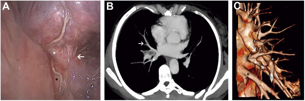
Commonly, V6 is the uppermost and smaller segmental tributary of the IPV.1 Nagashima et al. reported an uncommon drainage pattern in which V6 drained into the superior pulmonary vein.2 We describe a different variation: V6 draining into the MLV. Knowing this rare anatomical variant can help thoracic surgeons prevent intraoperative injuries during bilobectomies, right lower lobectomies and S6 segmentectomies. Careful examination of the vessels at preoperative CT-scan should be mandatory before starting any procedure.

Funding

None.

Conflict of interest

None declared.

References
[1]
D. Gossot, A. Seguin-Givelet.
Anatomical variations and pitfalls to know during thoracoscopic segmentectomies.
J Thorac Dis, 10 (2018), pp. S1134-S1144
[2]
T. Nagashima, K. Shimizu, Y. Ohtaki, K. Obayashi, S. Nakazawa, A. Mogi, et al.
Analysis of variation in bronchovascular pattern of the right middle and lower lobes of the lung using three-dimensional CT angiography and bronchography.
Gen Thorac Cardiovasc Surg, 65 (2017), pp. 343-349
Copyright © 2022. SEPAR
Archivos de Bronconeumología
Article options
Tools

Are you a health professional able to prescribe or dispense drugs?