Introduction
Obesity hypoventilation syndrome (OHS) is characterized by the existence of obesity and hypercapnic respiratory insufficiency which is not fully explained by neuromuscular, mechanical, or metabolic causes1. In fact, the causes of respiratory insufficiency and hypoventilation are not totally understood. A central origin has been suggested, either by itself or associated with the mechanical alterations of obesity on the respiratory system2. To date, obstructive sleep apnea syndrome (OSAS) has not been shown to play a causal role in the pathogenesis of OHS3. While there is evidence of the existence of a pure OHS, independent of OSAS, the two processes probably represent different evolutive forms of a single disorder in most cases4,5.
Noninvasive positive pressure ventilation (NPPV), which is usually applied at night, produces a clinical and functional improvement in OHS patients. Although the mechanism responsible for these effects may be multifactorial, the recovery of chemoreceptor sensitivity derived from improved gas exchange probably constitutes a very important factor6. One aspect that has not been studied in detail is the possibility of weaning this group of patients from ventilatory support once the respiratory insufficiency for which it was prescribed has improved. Withdrawal from positive pressure support has been shown to be associated with relapse in patients whose respiratory insufficiency is related to chest restriction7. However, the response to withdrawal may be different in patients with OHS, who have less marked changes in ventilatory mechanics, particularly if blood gases have been corrected with treatment and central ventilatory responses to chemical stimuli have been normalized. The aim of the present study was to evaluate the viability of withdrawing NPPV treatment from a group of patients with OHS.
Material and methods
Population studied
The patients included in the study had pure OHS and had received ventilatory support for a minimum period of one year. The study excluded patients who had other associated respiratory diseases as well as those who were treated with ventilatory support treatment for an average of less than 4 h/day. The initial diagnosis of OHS was made taking the following criteria into account: a) obesity, with a body mass index (BMI) greater than 30 kg/m²; b) diurnal and/or nocturnal hypercapnic respiratory insufficiency, with PaO2 measurements below 60 mmHg and PaCO2 measurements above 45 mmHg; c) lung function tests with a ratio of forced expiratory volume in 1 sec to forced vital capacity (FEV1/FVC) greater than 60%, and d) a nocturnal cardiorespiratory polygraphy with an apnea-hypopnea index (AHI) less than 10. All of the patients were receiving nocturnal ventilation from a bi-level positive airway pressure support device (BiPAP, Respironics Inc., Murrysville, PA, U.S.A.).
The criteria employed to evaluate the possibility of NPPV cessation were: a) a minimum time of one year since the first use of NPPV; b) lung function test with FEV1/FVC values greater than 60%; c) baseline arterial blood gases, both diurnal and nocturnal, with a PaO2 greater than 60 mmHg and a PaCO2 below 45 mmHg, and d) resting oxygen saturation greater than 90% for more than 70% of the night. The study excluded patients who did not meet these criteria. The study was approved by the hospital's ethics committee and all enrolled patients gave their informed consent.
Study design
An open, prospective study was carried out. Prior to withdrawing NPPV, we recorded nocturnal oximetry for all patients and determined awake and sleeping arterial blood gases. All patients also underwent lung function tests that included spirometry, body plethysmography, and ventilatory pattern studies, with basal occlusion pressure (P0.1) determination after hypercapnic stimulation. Patients who met the criteria for cessation of ventilation also underwent a nocturnal cardiorespiratory polygraph test. Three months after cessation of NPPV, all tests were repeated.
Diurnal arterial blood samples were extracted with the subject at rest and breathing air. The nighttime gases were extracted at 06:00 hours, with the patient awake. Both samples were processed in an ion-selective blood gas analyzer (IL-1306, Instrumentation Laboratory, Lexington, MA, U.S.A.), in accordance with the recommendations of the Spanish Society of Respiratory Diseases and Thoracic Surgery8. The nighttime oximetry was carried out on a portable apparatus (Ohmeda 4700 Oxicap, Louisville, Colorado, U.S.A.). Cardiorespiratory polygraphy was performed with a commercially available device (ApnoeScreen II+, Jaeger, GMBH, Wuerzburg, Germany) which had been previously validated in adults9. The spirometric and plethysmographic maneuvers were performed with the patient under sedation and with the nostrils occluded by a nose clip, following the recommendations of the European Respiratory Society10. Measurements were made using a MasterLab Pro (Jaeger, GMBH, Wuerzburg, Germany). P0.1 and breathing pattern were also determined with a MasterLab Pro apparatus (Jaeger, GMBH, Wuerzburg, Germany). To determine the ventilatory response to hypercapnia, the rebreathing technique was used as previously described by Read11.
Statistical analysis
The statistical analysis was performed using the SPSS 9.0 program for Windows. The descriptive data were expressed as means±standard deviations. To compare quantitative variables, nonparametric tests were used. Values of P<.05 were considered significant.
Results
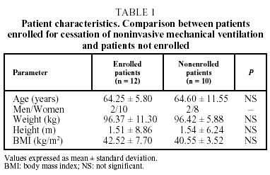
The study enrolled 22 patients with OHS who had received treatment with NPPV for a minimum period of one year. Of these, 12 (54.5%) met the criteria for withdrawal of NPPV while 10 (45.5%) did not. At the start of ventilation, there were no significant differences in patient characteristics or lung function tests between the two groups of patients. There were also no significant differences in patient weight between the times of starting and withdrawing ventilation. Finally, no differences between the groups were observed for patient characteristics at the time of evaluating cessation of NPPV (Table 1).
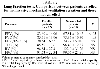
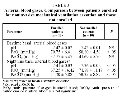
According to the established criteria, the lung function of patients from whom NPPV was withdrawn was normal or slightly below normal. All of the patients had PaO2 values higher than 60 mmHg and PaCO2 values lower than 45 mmHg. Patients who met all of the ventilatory support withdrawal criteria had FEV1 and FVC values (both parameters as a percentage of their predicted value) significantly higher than those of patients who could not have the treatment withdrawn (Table 2). Additionally, we saw differences in both daytime and nighttime PaO2 values and in daytime PaCO2 (Table 3). No significant differences between the two groups of patients were observed for oximetric variables, minimum saturation (84.0±4.0% for those from whom NPPV was withdrawn versus 73.0±18.2% for those who continued NPPV), or for the time during the night when oxygen saturation was greater than 90% (92.0±3.0% versus 88.0±5.4%, respectively).
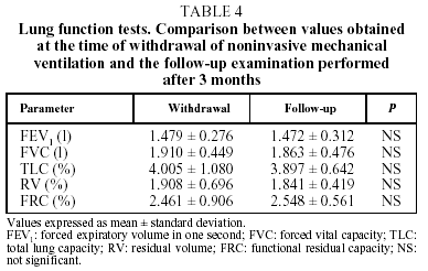
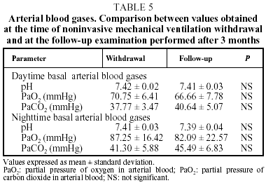
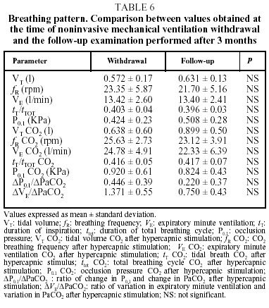
During the 3-month follow-up examination of the patients who were withdrawn from NPPV treatment, no significant changes were observed in weight (at follow-up: 99.4±16.4 kg), lung function test results (Table 4), daytime or nighttime arterial blood gases (Table 5), or oximetry (nighttime saturation parameters greater than 90% at the follow-up examination: 87.8±17.9%). There were no appreciable differences in breathing pattern or ventilatory response to hypercapnia. However, we did observe a tendency toward a diminished response to this chemical stimulus (Table 6).
In regard to the clinical course of individual patients in the study as revealed at a follow-up examination carried out 3 months later, only one had experienced a deterioration in symptoms and basal arterial gases that made it necessary to reintroduce NPPV. Finally, in 11 patients, 50% of those initially evaluated and 92% of those who were withdrawn from NPPV were able to remain free of ventilatory support.
In 7 of the 12 patients in the study (58.3%) the polygraph was consistent with OSAS. In fact, 4 of these patients had already showen signs of OSAS at the time of NPPV cessation (AHI: 11, 30, 57, and 66, respectively) and in 3 others OSAS became evident in the 3-month follow-up examination (AHI: 15, 21, and 62, respectively). These patients were included in our unit's OSAS group and treated with continuous positive airway pressure (CPAP). Finally, 4 patients remained in stable respiratory situations, showing no clinical deterioration and manifesting functional tests, blood gases, oximetry and cardiorespiratory polygraphs similar to those obtained at the time of liberation from ventilation.
Discussion
The present study demonstrates that, in a significant number of OHS patients who receive NPPV, this treatment can be interrupted in a relatively safe way, at least temporarily. In fact, 92% of the subjects who were withdrawn from this treatment were clinically stable 3 months later. Their lung function tests, blood gases, nighttime oximetry, breathing pattern, and response to hypercapnia were similar to those who were originally selected for liberation from NPPV.
NPPV has been shown to be efficient in the treatment of respiratory insufficiency originating in the CNS or in the chest wall. Although the causal mechanism is not totally defined, it has been postulated that nighttime ventilation could reverse daytime respiratory failure through an improvement in the sensitivity of the central chemoreceptors in the presence of chemical stimuli12. Nevertheless, the increase produced by this type of ventilation in lung volume, pulmonary distensibility, and the efficiency of respiratory musculature may be a contributing factor13.
The efficiency of NPPV treatment for OHS6,14,15 has been evaluated in only a few studies. In a recent controlled clinical trial its efficiency was compared to oxygen therapy in the treatment of obese patients with nocturnal hypoventilation and of subjects with chest wall disorders, fundamentally kyphoscoliosis16. In both these groups of patients a significant improvement was noted in the symptoms derived from nocturnal hypoventilation, with a rise in daytime PaO2 and a drop in nighttime PaCO2. But this improvement was seen only after application of noninvasive ventilation, with few modifications in the use of oxygen therapy, which to date has been considered the standard treatment. Later studies have confirmed that OHS patients improve clinically and functionally following treatment with noninvasive ventilation6. The level of improvement reached by OHS patients is similar to that obtained by patients with kyphoscoliosis, a disease in which NPPV has traditionally proven most effective.
There are few data on the repercussions of NPPV withdrawal, and those data that exist are derived mainly from the experience with patients with chest restriction17. Goldstein et al18 found that already on the first night without ventilation there was a worsening of nighttime oxygen saturation and PaCO2 in relation to the values recorded during ventilation. Hill et al19 later studied the repercussion of ventilatory support cessation in 6 patients with restrictive thoracic diseases treated with BiPAP. These authors detected worsening of symptoms after withdrawal, accompanied by a significant increase in the percentage of time at night with oxygen saturation of less than 90% and a decrease in minimum saturation but without significant changes in lung function, including basal arterial gases. These results are similar to those reported by Masa et al7, also in patients with thoracic disorders who were treated with NPPV. In this study, it was observed that withdrawal of ventilation for a period of 15 days produced significant alterations in the quality of sleep, with worsening nocturnal gas exchange and without modification of functional or muscle parameters. More recently, Karakurt et al20 have shown that interrupting NPPV for 6 days in patients with hypercapnic respiratory insufficiency, is accompanied by clinical and blood gas deterioration in 45% of patients. However, these authors evaluated not only patients with restrictive thoracic diseases, but also those with chronic obstructive pulmonary disease, the response to cessation being similar in both patient groups.
In OHS patients, the effect of cessation may be different because these patients present fewer mechanical changes than those with chest restriction. We observed no significant variations in the weight or BMI of patients who could be liberated from NPPV support in comparison with patients who could not be liberated. On the other hand, we did find differences in some blood gas and functional parameters whose values were higher in the former group of patients. The better functional situation of these subjects could explain, at least in part, why it proved possible to liberate them from ventilatory support. Another possibility would be that there exist differences in the respiratory driving behavior in the two groups of subjects. Although the design of this study does not permit evaluation of this aspect, because breathing pattern and ventilatory response to hypercapnia were studied only in those patients who met the criteria for withdrawal from NPPV, the evolution of our patients liberated from ventilation supports the possible relevance of differences in central respiratory driving. No significant changes were observed in weight or any of the lung function parameters in either group of patients, although we did see a tendency to worsening ventilatory response to chemical stimulation. The decrease in these patients' respiratory center sensitivity following the cessation of NPPV, while not statistically significant, can be interpreted as a possible initial sign of deterioration of the reconditioning which had theoretically been achieved with NPPV treatment. This could imply that after an undetermined period of time, longer than 3 months, the reset sensitivity of central chemoreceptors would progressively disappear. If this is the case, the progressive deficiency of the central respiratory signal would lead again to a condition of overall respiratory insufficiency like that originally manifested by these patients.
A high proportion of OHS patients developed OSAS when hypercapnia had been corrected. Of the 22 patients enrolled at the beginning of the study, 7 (32%) manifested a notable OSAS. Four of them were diagnosed at the time of withdrawing ventilation, and 3 were diagnosed at the follow-up examination. These patients were included in our unit's OSAS group and treated with CPAP. Several studies have shown that patients with moderate or acute OSAS treated with NPPV can sustain long-term absence of respiratory insufficiency if their ventilatory support treatment is followed by CPAP14,15. This explanation could justify the absence of deterioration in these patients after they are withdrawn from ventilation. In any case, the observed development allows us to propose that OHS is in fact one more phase of OSAS3. Perhaps central respiratory dysfunction and the hypoventilation to which it gives rise conceal an incipient clinical picture of OSAS. This condition would be manifested in this group of patients as OHS until the time when the resetting of central respiratory drive and the normalization of the upper airway inspiratory pressure flow rate led to the appearance of obstructive apneas. However, there is controversy about this and it is not clear whether OHS favors the development of OSAS or, to the contrary, the latter favors the development of the former in some patients. Additionally, it is possible that both are conditions that are aggravated by obesity and a variety of concomitant factors.
An obvious limitation to the study presented here is the small number of patients enrolled. It is prob able that a larger sample size would have made it possible to detect significant differences in some variables. Even so, the proportion of subjects we found able to do without mechanical ventilation 3 months after its cessation has, in and of itself, clinical relevance. Another limitation of the present study is the short follow-up period after NPPV withdrawal. Analysis after 3 months may only reflect an intermediate phase between the decrease in hypercapnic ventilatory response and the reappearance of respiratory insufficiency. This makes us consider the work we are presenting a preliminary study that requires a longer follow-up period to determine whether the withdrawal of ventilation can be maintained definitively or, to the contrary, whether all of the patients would end up returning irremediably to the situation they were in prior to beginning ventilation support.
Our conclusion, in accord with the results, is that NPPV can safely be suspended in some OHS patients, at least temporarily. These findings may shed light on the doubts that persist in relation to the physiopathology of this disease.
Correspondence: Dr. J. de Miguel Díez.
Servicio de Neumología. Hospital General Universitario Gregorio Marañón.
Dr. Esquerdo, 46. 28007 Madrid. Spain.
Manuscript received 30 October 2002.
Accepted for publication 26 November 2002.


