


A 59-year-old patient, with a history of right radical nephrectomy for clear cell renal cell carcinoma, presents in a control computed tomography (CT) scan (Fig. 1), a 14mm nodule in the posterior segment of the left lower lobe, not visible on previous CT scan, suspicious for lung metastasis.
 Axial CT section at T12 level in parenchyma window; (B) soft tissue window; (C) lung resection piece; (D) hyalinizing granuloma. In the center of the image, a foreign body (FB) of probable plant origin is observed, with calcified foci, encompassed by abundant fibrocollagenous tissue arranged in a laminar manner around it. In the periphery of the lesion, dilation of vascular spaces is identified, as well as a slight inflammatory reaction with the presence of lymphocytes and plasma cells.")
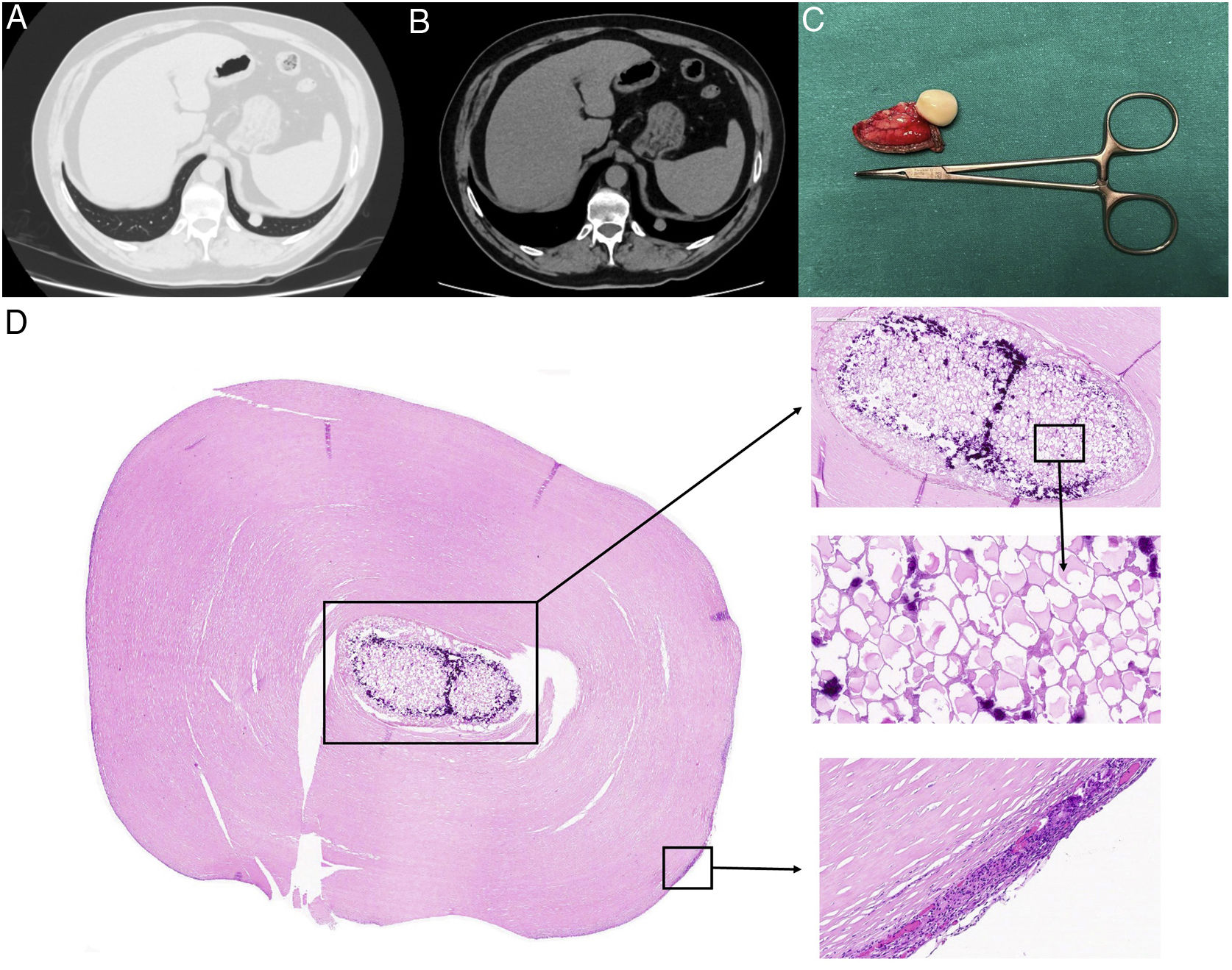
(A) Axial CT section at T12 level in parenchyma window; (B) soft tissue window; (C) lung resection piece; (D) hyalinizing granuloma. In the center of the image, a foreign body (FB) of probable plant origin is observed, with calcified foci, encompassed by abundant fibrocollagenous tissue arranged in a laminar manner around it. In the periphery of the lesion, dilation of vascular spaces is identified, as well as a slight inflammatory reaction with the presence of lymphocytes and plasma cells.
Wedge resection of the nodule is performed using video-assisted thoracoscopic surgery (VATS). The pathological anatomy result reports a hyalinizing granuloma, which includes material of probable plant origin (possible aspiration).
Intrathoracic foreign body (FB) are rare, but are usually present as a consequence of penetrating trauma or aspiration. Most of these FB occur in the bronchi, lungs, or esophagus, and there are few reports of intracardiac or intrapleural FB,1 as in this case.
The inflammatory response of the pleural space, to the stimulation of the FB, generates the formation of a hyalinizing granuloma,2 which in our case manifested as a pulmonary nodule, suggestive of metastasis.
Conflict of InterestThe authors declare that they have no conflict of interest related directly or indirectly to the contents of the manuscript.


