The unified airway hypothesis or global respiratory disease (GRD), is an established concept that the upper and lower airways form a single unified organ that is interconnected and interrelated by several physiologically important shared traits. These shared traits include immunology and pathophysiology, epidemiology, and clinical characteristics.
Accordingly, asthma shows a commonly comorbid conditions in the upper airways [1], most frequent rhinitis, allergic (AR) or non-allergic (NAR) and chronic rhinosinusitis (CRS) with (CRSwNP) or without (CRSsNP) nasal polyps. Their definitions have been exposed in the Allergic Rhinitis and its Impact on Asthma (ARIA) and the European Position Paper on Nasal Polyps and Sinusitis (EPOS) documents some time ago. Rhinitis, the Inflammation of the nasal mucosa is characterized by the following symptoms: nasal obstruction, rhinorrhea, sneezing and nasal pruritus/itching. It is often accompanied by ocular symptoms, such as pruritus and watery eyes. Two main phenotypes have been identified: (a) allergic rhinitis (AR), mediated by IgE after exposure to the allergen and skin prick tests and/or blood tests positive for inhaled allergens, and (b) non-allergic rhinitis (NAR), with negative allergy tests.
Chronic rhinosinusitis (CRS), the inflammation of the nasal mucosa and paranasal sinuses lasting over 12 weeks that manifests with 2 or more of the following symptoms, one of which must be congestion/obstruction/blocked nose and/or anterior/posterior rhinorrhea with or without the presence of pain/facial pressure and/or reduction/loss of sense of smell, and confirmed on nasal endoscopy and/or sinonasal computed tomography. There are two main phenotypes of CRS: CRSwNP and CRSsNP.
In asthma clinical practice, diagnostic delay of sinonasal comorbidities is common and has relevant consequences, on severity and control, particularly in CRSwNP associated with late-onset uncontrolled severe asthma.
Loss of smell, a key symptom associated with type 2 inflammation [2], is the only sinonasal symptom that significantly differentiates rhinitis from chronic rhinosinusitis and also distinguishes CRSwNP from CRSsNP. This finding was reported in a prospective real-life study [1] in 492 patients with asthma recruited from 23 centers in Spain and Latin America (IRIS-ASMA), where all sinonasal symptoms were evaluated according to ARIA and EPOS criteria using a visual analog scale (VAS, 0–100mm).
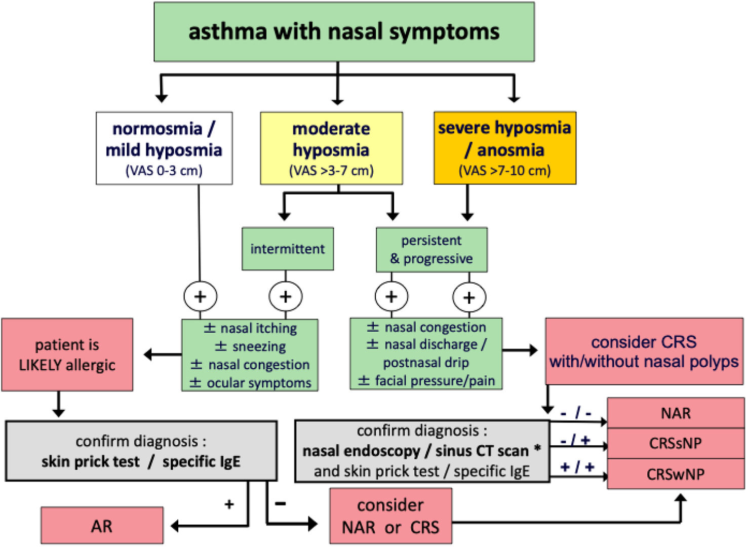
A loss of smell-based algorithm for the diagnosis of sinonasal comorbidities in asthma patients was first proposed in 2019 within a Sociedad Española de Neumología y Cirugía Torácica (SEPAR) asthma management document [3]. This algorithm guides the differential diagnosis of rhinitis vs. chronic rhinosinusitis comorbidities by using additional sinonasal symptoms and complementary diagnostic tests (skin prick test, specific IgE) and tools (nasal endoscopy, sinonasal CT scan).
This algorithm is used effectively in some Severe Asthma Units and Inflammation Rhinology Units in clinical practice in our hospitals, but since that publication [3], additional evidence has emerged, consistent with the role of inflammation in driving olfactory dysfunction, supporting its value in the diagnosis [2,4], severity assessment, and control of the disease, particularly in patients with CRSwNP. It has proven usefulness in the management of severe asthma and in guiding both the indication for and monitoring of response to biologic therapy, as highlighted in the POLINA [4], EPOS [5], and EUFOREA [6] recommendations. Newer biologic therapies have also been shown to improve olfactory function. Consequently, a loss of smell VAS score>7cm, considered severe hyposmia/anosmia, has been included among the criteria for initiating biologic treatment.
In addition, recent studies have validated the use of a VAS [7] for assessing olfactory loss, reinforcing the usefulness and reliability of this tool and justifying an update of the algorithm.
According to the EPOS [5] guidelines, loss of smell should be instrumentally assessed through validated smell tests (e.g., the University of Pennsylvania Smell Identification Test [UPSIT], Sniffin-Sticks, or the Barcelona Smell Test 24 [BAST-24]). However, other guidelines such as POLINA 2.0 [4] also suggest using a visual analog scale (VAS). As smell tests can be time consuming, an alternative tool to quickly assess which patients require a full olfactory work-up is necessary. There are two main options, either a short instrumental smell test or a subjective self-report.
The study population by Alobid I [7] et al. comprised 138 patients with severe CRSwNP and 40 controls. The BAST-24 identification score was strongly correlated with the VAS score in the CRSwNP group (p<0.001) but when VAS olfactory loss was categorized according to severity as 0–3cm, >3–7cm, and >7–10cm, a significant correlation was only demonstrated in the severe cases (VAS>7–10cm; p=0.002). In this study the best cut-off values for the VAS score was 5.2cm. The area under the receiver operating curve (AUC) was 0.85, with 72.5% sensitivity and 93.1% specificity.
With this new evidence [7], and by consensus among a multidisciplinary group (two pulmonologists, two allergists, and four otorhinolaryngologists), we present an updated version of this diagnostic algorithm (see Fig. 1).

Diagnostic algorithm for patients with asthma and sinonasal symptoms to differentiate, according to the severity of olfactory loss (normosmia, hyposmia and anosmia), between rhinitis (allergic or non-allergic) and chronic rhinosinusitis with or without nasal polyps. In cases of partial (hyposmia) or complete (anosmia) loss of smell, other common causes of olfactory dysfunction should first be ruled out, such as previous head trauma, recent upper respiratory tract infection (e.g., common cold, influenza, or COVID-19), or neurodegenerative disease (e.g., Parkinson's disease or Alzheimer's disease). Abbreviations: CRSsNP, chronic rhinosinusitis without nasal polyps; CRSwNP, chronic rhinosinusitis with nasal polyps; AR, allergic rhinitis, NAR, non-allergic rhinitis; VAS, visual analog scale (0–10cm). (*) Recommended only after nasal endoscopy if needed or presurgical or for diagnosis purposes only, sinonasal CT scan is recommended to be done under a low radiation protocol.
This multidisciplinary group experts on asthma and CRSwNP, from different centers met with the aim of developing a symptom test, included the decrease or loss of smell, had to be easy to perform and implemented, such as visual analog scale-based PRO (patient reported outcome) that could be completed in the waiting room or at the consultation for interdisciplinary use for the management of GRD, that help the physician in the taking decisions for diagnosis and treatment of upper airways comorbidities in asthma patients.
The updated algorithm was developed by this group of otorhinolaryngologists, pulmonologists and allergists with expertise in rhinology through a literature-informed process, synthesizing recurrent clinical–inflammatory patterns described in previous pheno/endotype classifications, cluster analyses, and consensus documents. Although not conducted as a formal Delphi process, iterative structured discussions were performed to ensure conceptual coherence and alignment with existing evidence.
Consensus was defined by as more than >70% agreement of authors. Each step and cut-off points in the algorithm is grounded on a consensus, after three rounds of review in which the discussion focused on the decisions to diagnosis based on clinical symptoms and issues to the standard diagnostic strategy [3]. The 4-step diagnostic algorithm proposed in the latest SEPAR guidelines document [3] for the management of global respiratory disease was considered, with special mention that sinonasal CT scan is recommended only after nasal endoscopy if needed or for diagnostic purpose or presurgical, and recommended to be done under a low radiation protocol.
Panelists reached consensus on, and hence recognized, the importance of testing the reduction in or loss of smell [1,8–10]. The updated algorithm adds the VAS cut-off points into normosmia (0–3cm), hyposmia>3–7cm, and severe hyposmia/anosmia (>7cm), using a 0–10cm VAS has been defined by the POLINA [4] guideline.
Although CRS is the most frequent cause of olfactory dysfunction (OD), mainly CRSwNP but also CRSsNP, other causes of OD must be investigated.
So, in cases of partial (hyposmia) or complete (anosmia) loss of smell, other common causes of olfactory dysfunction should first be ruled out, such as immunosuppressive medication, previous head trauma, prior nasal surgery, recent upper respiratory tract infection (e.g., common cold, influenza, or COVID-19), or neurodegenerative disease (e.g., Parkinson's disease, dementia or Alzheimer's disease), aging-related hyposmia (presbyosmia) or systemic inflammatory disease, before considering loss of smell in a patient evaluation.
There is a clear need for simple and practical tools, that help clinicians to diagnose severe asthma and its comorbidities such as CRSwNP [8].
The resulted evidence based algorithm is easy to use in current clinical practice and may help both specialists and primary care physicians to reduce the diagnostic delay [8] and initiate the appropriate treatment of sinonasal asthma comorbidities. In addition, this symptom-based smell tool would be useful in epidemiological studies and in different clinical settings including specialists and primary care physicians where endoscopic or imaging outcomes are not available or even considered.
This updated diagnostic algorithm will need to be validated in clinical practice and further prospective studies are needed to confirm its impact on reducing underdiagnosis and improving clinical outcomes.
Author contributionJAC and JM projected and performed the original study and designed the algorithm, IA, EA, IB, JD, MG and SS all contributed to the analysis of this tool. JAC drafted the paper and all authors have reviewed and edited the paper and approved the final version.
Artificial intelligence involvementThe authors used ChatGPT (Open AI; GPT-5.3) for English language editing. All content was reviewed and approved by the authors.
FundingThis research received no specific grant from any funding agency in the public, commercial, or not-for-profit sectors.
Conflict of interestJAC has received research grants, attended speaker bureaus and/or advisory boards from ALK-Abelló, AstraZeneca, GlaxoSmithKline, MSD, and Sanofi-Regeneron.
IA has received honoraria for consultancy and conferences from AstraZeneca, Viatris, Roche, Sanofi, GSK, MSD, Menarini, Storz, Olympus, Metronic, Salvat and Novartis.
EA has received fees in the past three years as speaker, scientific advisor,and/or clinical investigator from AstraZeneca, Chiesi, Gebro, GlaxoSmithKline, Menarini, MSD, Orion Pharma, and Sanofi-Regeneron.
IB has received honoraria for consultancy and conferences from AstraZeneca, Sanofi-Regeneron, GSK, Menarini, Chiesi, Novartis.
JDO has received fees in the past three years as a consultant from AstraZeneca, GSK and Sanofi-Regeneron and as a speaker at meetings sponsored by ALK-Abelló, AstraZeneca, Chiesi, GlaxoSmithKline, LETI, Novartis, Mundipharma, Sanofi-Regeneron, Stallergenes, and TEVA.
MG has participated in advisory boards and speaker activities and has received consulting fees from AstraZeneca, GSK, and Sanofi-Regeneron.
SS has received honoraria for consultancy and conferences from AstraZeneca, Sanofi, GSK, and Medtronic.
JM has received research grants, attended speaker bureaus and/or advisory boards and received consulting fees from Almirall, AstraZeneca, Berlin-Chemie, GSK, Eli Lilly, Menarini, Mitsubishi-Tanabe Pharma, MSD, Noucor/Uriach Group, Novartis, Regeneron Pharmaceuticals Inc., and Sanofi-Genzyme, UCB Pharma, and Viatris.