


A 49-year-old man with stage IV sarcoidosis presented with worsening respiratory symptoms and low-grade fever.
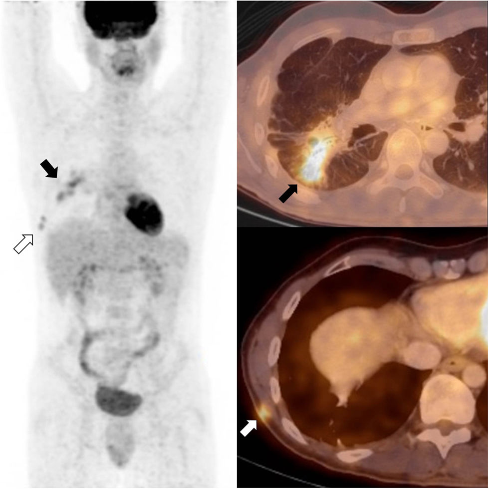
18FDG-PET-CT (fig. 1) showed confluent hypermetabolic nodules in the right lower lobe (RLL) consistent with reactivation of pulmonary sarcoidosis (black arrows). Besides, two small hypermetabolic subcutaneous nodules were revealed in the right thoracic wall. After carefully reviewing the medical record and the previous CT images, we arrived at the diagnosis of scar sarcoidosis at the scar of a video-assisted thoracoscopic biopsy of the RLL performed 10 years before, during the initial diagnostic work-up (white arrows). Treatment with infliximab was initiated and both the pulmonary and the cutaneous lesions resolved completely, thus confirming the diagnosis.Cutaneous sarcoidosis has many forms and affects 25% of patients with sarcoidosis. Scar sarcoidosis, an uncommon form of cutaneous sarcoidosis, represents less than 3% of the cases of skin involvement1. It consists in the development of sarcoid granulomas on previous scar tissue, and it should not be mistaken with keloids.
 and two small hypermetabolic subcutaneous nodules in the right thoracic wall (white arrows).")
18FDG -PET-CT is an emerging tool for the staging and response assessment of sarcoidosis2. In this case, the high sensibility of 18FDG-PET was key to detect the granulomas, whereas the spatial and contrast resolution of the CT allowed the precise characterization of their location and nature.


