Alpha-1 antitrypsin deficiency (AATD) is a rare inherited disorder caused by mutations in the SERPINA1 gene. AATD is one of the leading causes of emphysema in young adults and of liver cirrhosis in children and adults. Despite significant underdiagnosis and high clinical variability, recent research is rapidly transforming the field. Advances in molecular biology and translational medicine have identified new biomarkers and therapeutic targets, although reliable predictors of disease progression remain limited. While augmentation therapy is the only approved pharmacological option for pulmonary involvement and no specific treatment exists for liver disease, innovative strategies are remodeling the therapeutic landscape. Gene therapy, RNA interference, gene editing, and epigenetic modulation are showing promising preclinical and early clinical results, aiming to correct the underlying defect rather than manage symptoms. This review focuses on these emerging advances, highlighting cutting-edge preclinical and translational developments with potential to redefine AATD management and move toward curative approaches.
Alpha-1 antitrypsin deficiency (AATD) (ORPHA: 60) is a rare hereditary disorder (prevalence of 1–5:10.000), caused by mutations in the SERPINA1 gene. The Pi*Z (Glu342Lys) allele is the most clinically significant. This variant leads to abnormal folding of the alpha-1 antitrypsin (AAT) protein, promoting its polymerization and intracellular accumulation within the endoplasmic reticulum (ER) of hepatocytes (as well as monocytes, neutrophils and epithelial cells) [1,2]. As a result, the disease has a dual pathogenesis. The accumulation of AAT polymers in the liver can lead to liver fibrosis and cirrhosis development, whereas systemic deficiency of functional AAT promotes a protease-antiprotease imbalance in the lung leading to parenchymal destruction and the development of emphysema [1,3–5].
AATD patients face three main challenges: underdiagnosis, high clinical variability and lack of specific pharmacological treatments for liver disease. More than 90% of cases remain undiagnosed. Initial nonspecific symptoms make early diagnosis difficult and leads to delays of up to 5–10 years, which negatively impacts the prognosis. However, we would like to point out that the frequently cited estimate that up to 90% of AATD cases remain undiagnosed is subject to considerable uncertainty [6]. This figure is derived by comparing expected prevalence, estimated from population screening and SERPINA1 allele frequencies, with documented cases, and may vary according to geographical region, genotype, and diagnostic strategy [7]. Clinical severity varies widely, and neither serum AAT levels nor the phenotype reliably predicts disease progression, suggesting the influence of genetic, epigenetic, environmental, and lifestyle factors [4,5]. Current research focuses on early diagnostic strategies [8], and discovery of prognostic biomarkers. Augmentation therapy is the only approved pharmacologic treatment for pulmonary disease, although its uptake has been variable across countries, mainly due to differences in cost and local healthcare economics, augmentation therapy is endorsed by current guidelines based on the available scientific evidence. Finally, there is no specific treatment for liver disease, although new specific treatments based on gene therapy technologies are already in the clinical trial phase and can change the paradigm of the disease [9–11].
Given the extensive literature already available on AATD, this review focuses on the most recent advances, with a particular emphasis on preclinical and translational results not yet implemented in clinical practice, while also considering the role of traditional therapeutic approaches within a comprehensive approach.
Molecular mechanisms with therapeutic relevanceEndoplasmic reticulum stress and unfolded protein responseAs mentioned above, AATD is caused by the abnormal folding of the AAT Z variant, which promotes its polymerization and intracellular retention, constituting the initial event in the pathogenesis [1,11].
The accumulation of misfolded AAT in the ER causes cellular stress and activates the unfolded protein response (UPR), a protective mechanism aimed at restoring cellular balance. However, when this activation is chronic, it contributes to inflammation, cell death, and progression of liver damage. At the same time, reduced levels of functional AAT in the circulation disrupt the protease–antiprotease balance in the lung, allowing enzymes such as neutrophil elastase to damage lung tissue. Cells normally remove abnormal proteins through quality control systems, such as endoplasmic reticulum-associated degradation (ERAD), as well as proteasomal degradation and autophagy. When these mechanisms are overwhelmed or impaired, misfolded proteins accumulate further, worsening tissue injury [12–15].
These processes represent important therapeutic targets. Emerging strategies such as protein folding modulators, autophagy inducers, polymerization inhibitors, and gene therapies targeting SERPINA1 aim to directly intervene in the underlying molecular mechanisms, with the goal of correcting both hepatic toxicity and systemic deficiency which will be further discussed ahead.
Genetic modifications and variable phenotypeSERPINA1 is a highly polymorphic gene with more than 500 variants described to date [16], exhibiting a broad spectrum of pathogenicity ranging from benign to disease-causing. An ever-increasing spectrum of SERPINA1 mutations has recently been identified, giving rise to AAT variants associated with deficiency and/or dysfunctional behavior [17,18].
Pathogenic AAT variants are classified as null or deficient. Null variants (Q0) are characterized by lack of AAT production, leading to undetectable serum levels and a high risk of early-onset emphysema, even in non-smokers [19]. Deficient variants are caused by point mutations causing intracellular retention, polymer formation, increased degradation, or reduced antiprotease activity. These mechanisms are not mutually exclusive producing variable degrees of quantitative and functional deficiency. For example, the Z allele, the most common severe variant, is highly polymerogenic and characterized by marked intracellular retention, although the small fraction that is secreted retains residual, but impaired, inhibitory activity against neutrophil elastase [20]. In contrast, the rare F variant is non-polymerogenic, functionally impaired, and only mildly reduced in circulating levels [21].
Genetic mutations alone, or in combination with environmental factors such as cigarette smoke, which is known to induce post-translational modifications including oxidation or polymerization [22], may disrupt AAT trafficking in pulmonary endothelial cells, including its cellular uptake, intracellular transport, and secretion, thereby affecting its functional bioavailability within specific lung compartments [23].
The diverse pathogenic mechanisms associated with AAT variants, which are expressed in a codominant fashion and can therefore result in a wide range of genotypic combinations [24], represent the phenotypic consequences of the heterogeneity in the clinical presentation of AATD patients.
Recent spatial and genomic studies have refined our understanding of AATD-associated liver disease. The spatial study recapitulates known markers of disease progression and identifies dysregulated proteins. A key finding is the early activation of the peroxisomal response in patients with low-grade liver fibrosis, whereas this response is delayed in advanced fibrosis. Because this association is not observed in alcohol-related liver disease, it may represent a mechanism specific to AATD [25]. Analysis of somatic mutations has shown that chronic Z-AAT proteotoxicity influences hepatocyte clonal evolution, favoring the expansion of clones with acquired alterations that reduce mutant AAT production or its intracellular burden [26].
Advances in AATD diagnosisClinical diagnosisAATD clinical diagnosis is challenging due to phenotypic heterogeneity and the lack of pathognomonic symptoms [27,28]. Contrary to the typical AATD patient profile of a young (under 50 years of age at the time of diagnosis), nonsmoker individual with basal panacinar emphysema, over 50% of cases are diagnosed after age 50 [29–31], around 36% of patients present apical emphysema [32], and approximately 80% are current smokers (8%) or former smokers (71%) [32–34].
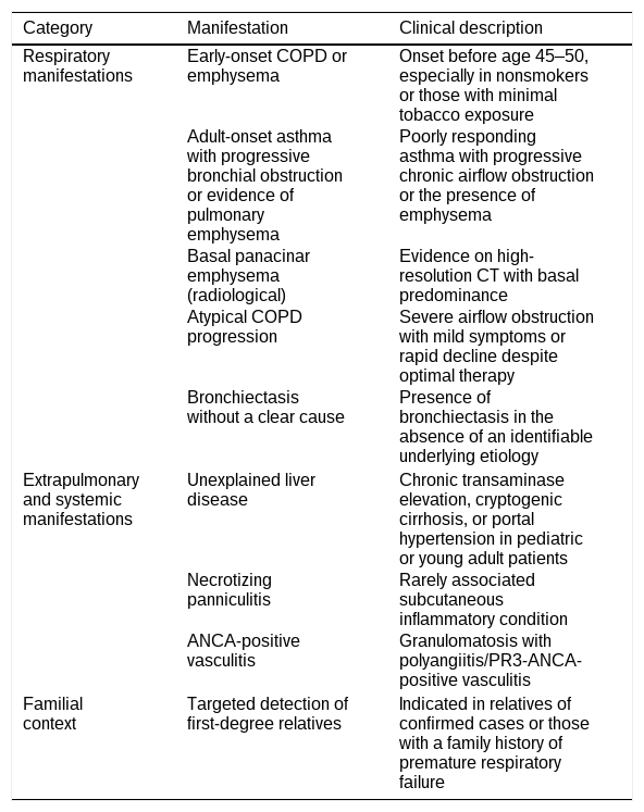
The diagnostic algorithm must be initiated upon detection of specific pulmonary, hepatic, or systemic “red flags” (Table 1) [35]. According to the ATS/ERS statement, AATD testing is recommended in individuals with COPD or otherwise unexplained persistent airflow obstruction, unexplained chronic liver disease, necrotizing panniculitis, and unexplained bronchiectasis [6,28]. These documents also specifically mention granulomatosis with polyangiitis/PR3-ANCA-positive vasculitis. However, this indication should not be generalized to all ANCA-associated vasculitis [36]. Genetic counseling and targeted testing should be offered to first-degree relatives of individuals with a confirmed deficiency-associated SERPINA1 variant. A family history of premature severe COPD may increase clinical suspicion but is not, by itself, an indication for familial testing [4,6,37,38].
Clinical indicators for AATD screening.
| Category | Manifestation | Clinical description |
|---|---|---|
| Respiratory manifestations | Early-onset COPD or emphysema | Onset before age 45–50, especially in nonsmokers or those with minimal tobacco exposure |
| Adult-onset asthma with progressive bronchial obstruction or evidence of pulmonary emphysema | Poorly responding asthma with progressive chronic airflow obstruction or the presence of emphysema | |
| Basal panacinar emphysema (radiological) | Evidence on high-resolution CT with basal predominance | |
| Atypical COPD progression | Severe airflow obstruction with mild symptoms or rapid decline despite optimal therapy | |
| Bronchiectasis without a clear cause | Presence of bronchiectasis in the absence of an identifiable underlying etiology | |
| Extrapulmonary and systemic manifestations | Unexplained liver disease | Chronic transaminase elevation, cryptogenic cirrhosis, or portal hypertension in pediatric or young adult patients |
| Necrotizing panniculitis | Rarely associated subcutaneous inflammatory condition | |
| ANCA-positive vasculitis | Granulomatosis with polyangiitis/PR3-ANCA-positive vasculitis | |
| Familial context | Targeted detection of first-degree relatives | Indicated in relatives of confirmed cases or those with a family history of premature respiratory failure |
The most significant advance has been implementing active case-finding programs and diagnostic algorithms in primary care combining clinical criteria with initial laboratory tests to facilitate early detection [8,39].
Molecular diagnosisDespite the success of active case-finding and primary care algorithms in improving early detection, clinical and radiological criteria alone cannot confirm the disease. A definitive diagnosis requires the use of comprehensive molecular strategies, including SERPINA1 genotyping and sequencing. Although there is no universally accepted diagnostic algorithm, a widely accepted one has been proposed [5,8,35]. The diagnostic approach includes: (i) measurement of plasma AAT levels, (ii) protein phenotyping, (iii) genotyping targeting the most common variants [28], and (iv) full sequencing in those cases with low AAT serum levels but no abnormal allele is found by PCR or when null or rare variants are suspected [40–42].
Overall, these molecular techniques are needed to identify rare alleles or null variants improving diagnostic accuracy where rapid methods fail [43–45]. In addition, the development of high-throughput platforms allows familial and targeted population detection in at-risk populations. Importantly, precision molecular biology-based approaches are being developed that correlate genotype with clinical risk, enabling better patient stratification [5].
Biomarkers in AATDBiomarkers proposed for use in AATD differ substantially in their intended purpose. Some serve a diagnostic role (confirming deficiency or genotype), others are pharmacodynamic (demonstrating biochemical exposure to treatment), and others are proposed as markers of ongoing disease activity or progression. However, currently, no single circulating biomarker is specific and validated enough to measure pulmonary disease activity or emphysema progression in AATD.
Biomarkers of progressionIdentifying reliable biomarkers for monitoring the AATD progression is an area of intense research. From a clinical point of view, it is necessary to identify biomarkers that help identify patients with a poorer prognosis.
Limitations of FEV1 as a reference standardRegarding lung function, spirometry (particularly FEV1) remains the main tool for assessing pulmonary progression. However, FEV1 is a poor surrogate for AATD-related emphysema, as it insensitively reflects cumulative, late-stage parenchymal destruction [32,46]. Quantitative CT densitometry detects progression and treatment effects with greater sensitivity. Therefore, biomarker validation in AATD should rely on longitudinal CT densitometry and complementary outcomes rather than cross-sectional FEV1 correlation alone [47,48].
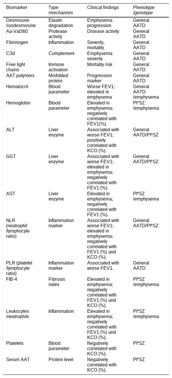
Various biomarkers have been studied to assess lung damage and the progression of emphysema in AATD. The correlation between serum biomarkers and pulmonary function is shown in Table 2.
Lung biomarkers in AATD.
| Biomarker | Type/mechanism | Clinical findings | Phenotype/genotype |
|---|---|---|---|
| Desmosine/isodesmosine | Elastin degradation | Emphysema progression | General AATD |
| Aa-Val360 | Protease activity | Disease activity | General AATD |
| Fibrinogen | Inflammation | Severity, mortality | General AATD |
| C3d | Complement | Emphysema severity | General AATD |
| Free light chains | Immune activation | Mortality risk | General AATD |
| AAT polymers | Misfolded protein | Progression marker | General AATD |
| Hematocrit | Blood parameter | Worse FEV1; elevated in emphysema | General AATD/emphysema |
| Hemoglobin | Blood parameter | Elevated in emphysema; negatively correlated with FEV1(%). | PI*SZ/emphysema |
| ALT | Liver enzyme | Associated with worse FEV1; positively correlated with KCO (%). | General AATD/PI*SZ |
| GGT | Liver enzyme | Associated with worse FEV1; elevated in emphysema; negatively correlated with FEV1 (%). | General AATD/PI*SZ |
| AST | Liver enzyme | Elevated in emphysema; negatively correlated with FEV1 (%). | PI*SZ/emphysema |
| NLR (neutrophil/lymphocyte ratio) | Inflammation marker | Associated with worse FEV1; elevated in emphysema; negatively correlated with FEV1 (%) and KCO (%). | General AATD/PI*SZ |
| PLR (platelet/lymphocyte ratio) | Inflammation marker | Associated with worse FEV1. | General AATD |
| FIB-4 | Fibrosis index | Elevated in emphysema; negatively correlated with FEV1 (%) and KCO (%). | PI*SZ/emphysema |
| Leukocytes/neutrophils | Inflammation | Elevated in emphysema; negatively correlated with FEV1 (%) and KCO (%). | PI*SZ/emphysema |
| Platelets | Blood parameter | Negatively correlated with KCO (%). | PI*SZ |
| Serum AAT | Protein level | Negatively correlated with KCO (%). | PI*SZ |
Desmosine and isodesmosine are released during elastin degradation and have been investigated as potential biomarkers of tissue damage in COPD and AATD [49]. Associations with impaired lung function, radiological emphysema, and clinical outcomes have been reported in some cohorts [50,51]. However, circulating and urinary levels are not specific to lung tissue, and findings regarding their ability to predict longitudinal emphysema progression have been inconsistent [52]. Their principal limitation is lack of organ specificity, since elastin is also present in vasculature and skin; levels are additionally affected by age, cardiovascular disease, and renal clearance [49,50].
Neutrophil elastase activity markers and Aa-Val360Because AATD-related emphysema results from an imbalance between neutrophil elastase and its principal inhibitor, direct assessment of elastase activity (either in sputum/BAL or via the fibrinogen-derived Aa-Val360) has strong mechanistic rationale [53,54]. Nevertheless, sputum and BAL measurements are affected by sampling technique, dilution, and airway infection, and BAL is invasive and unsuitable for repeated sampling.
Aa-Val360 is elevated circulating levels and association with markers of disease severity and reductions after intravenous augmentation therapy have been reported [55]. However, the available evidence is based on relatively small cohorts, and Aa-Val360 has not been validated as a marker of clinical disease activity or emphysema severity and its ability to predict longitudinal lung-function decline or CT lung-density loss remains to be established [56]. Aa-Val360, while more specific to elastase-mediated proteolysis, is not lung-specific and may be influenced by systemic neutrophilic inflammation [53,54].
Systemic inflammatory markersThese markers have prognostic value in COPD in general (57,58). However, none is specific to AATD; all are influenced by smoking, obesity, infections, exacerbations, and cardiovascular or metabolic comorbidities [57–59]. In this sense, extrapolation of findings from usual COPD to AATD should be made cautiously, as the underlying pathophysiology and typical age of onset differ.
Systemic inflammatory biomarkers like c-reactive protein (CRP) and CCL18 reflect the chronic inflammation associated with AATD-related lung damage, with significantly higher levels in patients with emphysema.
Aljama et al. [60] showed that higher hematocrit, elevated ALT and GGT levels, and a higher neutrophil-to-lymphocyte ratio (NLR) and platelet-to-lymphocyte ratio were associated with a lower FEV1. Lopez-Gonzalez et al. found that patients with emphysema had elevated levels of AST, GGT, FIB-4 index, hemoglobin, leukocytes, neutrophils, and NLR. In PI*SZ patients, FEV1 showed weak but significant negative correlations with these biomarkers, while KCO was negatively correlated with AAT, FIB-4, leukocytes, neutrophils, platelets, and NLR, and positively correlated with ALT [61].
Epithelial-derived biomarkers (CC16)These proteins originate largely from the lung and have been studied as markers of epithelial injury or increased alveolar-capillary permeability. Despite their pulmonary origin, they are not disease-specific and while CC16 has been reported to predict emphysema progression in COPD patients [62], its usefulness is limited since its levels are affected by smoking status, age, and renal clearance [62,63].
More recently, proteomic approaches have identified novel molecular signatures associated with lung function decline [64].
However, the use of serum biomarkers to assess disease severity should be approached with caution, as weak correlations make it difficult to identify a single reliable biomarker for accurately predicting disease severity and progression.
Liver damage in AATD is caused by intrahepatic accumulation of misfolded AAT polymers, leading to cellular stress, inflammation, and fibrosis. Non-invasive methods are increasingly used to detect and monitor liver involvement in AATD, as routine liver enzyme levels may remain normal despite clinically relevant fibrosis [65–68]. Serum-based fibrosis scores, such as APRI and FIB-4, combined with liver stiffness measurement by transient elastography, may facilitate risk stratification and identify patients requiring closer monitoring or further diagnostic evaluation [65]. However, liver stiffness can be influenced by inflammation, cholestasis, congestion, food intake and technical factors, while APRI and FIB-4 have not been as extensively validated in AATD as in other chronic liver diseases.
Recent consensus evidence supports the use of transient elastography use for detecting and stratifying clinically relevant fibrosis. Therefore, liver stiffness measurement should be incorporated into the liver evaluation of patients with AATD, particularly those with the Pi*ZZ genotype [69–71].
In summary, current recommendations support an integrated assessment combining clinical findings, routine laboratory tests, platelet count, serum fibrosis scores, liver imaging and liver stiffness measurement [68–71].
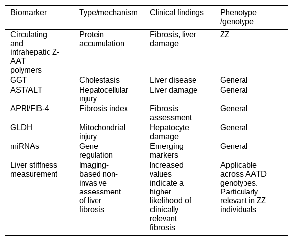
Conventional liver tests lack sensitivity, but biomarkers such as circulating AAT polymers, fibrosis indices, and emerging molecular markers may improve early detection and monitoring disease progression (Table 3).
Hepatic biomarkers in AATD.
| Biomarker | Type/mechanism | Clinical findings | Phenotype/genotype |
|---|---|---|---|
| Circulating and intrahepatic Z-AAT polymers | Protein accumulation | Fibrosis, liver damage | ZZ |
| GGT | Cholestasis | Liver disease | General |
| AST/ALT | Hepatocellular injury | Liver damage | General |
| APRI/FIB-4 | Fibrosis index | Fibrosis assessment | General |
| GLDH | Mitochondrial injury | Hepatocyte damage | General |
| miRNAs | Gene regulation | Emerging markers | General |
| Liver stiffness measurement | Imaging-based non-invasive assessment of liver fibrosis | Increased values indicate a higher likelihood of clinically relevant fibrosis | Applicable across AATD genotypes. Particularly relevant in ZZ individuals |
Circulating AAT polymers reflect intrahepatic accumulation of abnormal protein and correlate with liver dysfunction and disease severity. Their plasma levels are associated with GGT and other biochemical markers of liver injury, and higher concentrations are associated with increased liver stiffness and the presence of liver disease [55,72]. At the tissue level, liver AAT polymer load is directly associated with fibrosis progression and adverse outcomes, including liver-related mortality, supporting their role as both diagnostic and disease progression markers [73]. Furthermore, recent studies show that serum AAT polymer levels and hepatic accumulation are associated with advanced fibrosis stages and correlate with AST and the Aspartate Aminotransferase-to-Platelet Ratio Index (APRI score), supporting their value as biomarkers of structural liver damage [74].
The intrahepatic retention of misfolded and polymerized Z-AAT is central to the pathogenesis of AATD-associated liver disease [75,76]. Clark et al. demonstrated an association between intrahepatic AAT accumulation and histological liver abnormalities, including fibrosis, in adults with the Pi*ZZ genotype [77]. More recently, Fromme et al. showed that circulating Z-polymer levels were associated with liver fibrosis and adverse clinical outcomes in adults with AATD [78]. These findings support the potential value of circulating Z-polymers as non-invasive prognostic biomarkers; however, further assay standardization and prospective validation are required before their routine clinical implementation.
In addition, new biomarkers derived directly from AAT, such as C-terminal peptides, are currently being investigated. In a pediatric study, C37 and C42 have been shown to correlate with transaminases (ALT and AST) and may reflect early hepatocellular damage [79].
Advances in AATD. Development of new therapeutic strategiesPreclinical modelsPreclinical models of AATD, including cell cultures, and animal and human models have been essential for understanding disease mechanisms and developing new therapies.
Common cellular models include HEK293, COS-7, and CHO cells engineered to express mutant AAT variants, especially the Z allele, and are used to study polymerization, ER retention, and degradation pathways such as ER-associated degradation and autophagy [80–82]. Hepatocyte-derived cell lines such as HepG2 and Huh7 hepatoma-derived cells provide a more physiological hepatic context and are widely used to analyze secretion defects, polymer accumulation, ER stress, and pharmacological responses [83,84]. These systems are valued for high transfection efficiency, strong protein expression, and suitability for proteostasis and therapeutic screening.
More advanced cellular models include patient-derived iPSCs that has been differentiated into hepatocyte-like cells [85], which preserve patient genetic background and reproduce intracellular polymer accumulation, making them highly valuable for precision medicine and gene-editing validation [86–88]. In AATD-patient-derived iPSCs, the correction of Z variant can restore the structure and function of AAT [86].
Additionally, three-dimensional liver organoids recapitulated the key features of Z-AAT deficiency [89,90]. Liver organoid models derived from iPSCs, primary cells and from liver biopsies of patients with specific genotypes (i.e. ZZ, MZ) are being very useful for investigating responses and pathogenic features enabling an improved tissue architecture, and long-term modelling [91,92].
Animal models are essential for studying systemic AATD. PiZ transgenic mice (C57BL/6-PiZ and NSG-PiZ), expressing human Z-AAT, are widely used for liver disease and show hepatocellular polymer accumulation, ER stress, and progressive liver injury [81,93,94]. However, PiZ mice do not develop spontaneous emphysema, since murine neutrophil elastase is not efficiently inhibited by human Z-AAT, and mice lack an endogenous protease–antiprotease imbalance comparable to that in humans [93,95]. This model is therefore useful for hepatic but not pulmonary disease. In addition, the Serpina1 a–e knockout mice lack endogenous AAT and model pulmonary disease with protease–antiprotease imbalance and emphysema-like damage. Limitations include the absence of the toxic gain-of-function polymer accumulation seen with the Z variant, meaning this model isolates loss-of-function effects but does not recapitulate the combined mechanisms operating in human PiZZ disease [96]. Recently, a new AAT-null PiZ strain combines SERPINA1 knockout with human PiZ expression [97].
More recent approaches have generated mice that both lack murine AAT and express human Z-AAT, aiming to combine loss-of-function and gain-of-function mechanisms in a single model. CRISPR/Cas9-based gene editing has also been used to generate more precise humanized models and to explore gene-correction strategies as potential therapeutic approaches. These models remain relatively new, and long-term data on their ability to reproduce progressive emphysema and their utility for therapeutic screening are still limited [98,99].
Gene therapy and RNA-based therapiesDNA editing: permanent correctionThe evolution of gene therapy for AATD arises from already classic studies that established the proof of concept for hepatic gene transfer. Pioneering research by Aliño et al. first demonstrated the feasibility of using non-viral vectors and liposomes to deliver the SERPINA1 gene in animal models [100,101]. Further studies by this group, explored hydrodynamic injection to achieve sustained expression of human AAT protein within the liver parenchyma [102,103].
Currently, the field has advanced toward correcting the SERPINA1 locus using adenine base editors (ABEs). Preclinical studies using lipid nanoparticles achieved 35% editing efficiency in hepatocytes, raising serum AAT above the therapeutic threshold of 11μM and reducing the intrahepatic polymers [104]. Additionally, CRISPR/Cas9 via AAV8 vectors deleted the Z allele, reducing fibrosis and inflammation [98]. These approaches represent a shift from augmentation therapy to a potential one-time curative treatment by addressing the genetic cause in both the liver and the lungs. Recently, it has been shown that ABE-based correction of the Z variant in patient iPSCs reduces aberrant AAT accumulation and restores its secretion [91,105]. Finally, ABE editing has recently entered clinical development for AATD. BEAM-302 is a liver-targeted adenine base editor designed to correct the pathogenic SERPINA1 Z variant without generating double-strand DNA breaks and is currently being evaluated in a first-in-human phase 1/2 trial (NCT06389877). Preliminary findings have shown the appearance of corrected M-AAT, increased total and functional AAT levels, and reduced circulating Z-AAT, providing initial clinical proof of mechanism. Nevertheless, the BEAM-302 findings remain preliminary, and longer follow-up and larger cohorts are required to establish safety, editing durability, and effects on pulmonary and hepatic clinical outcomes.
Altogether, these results demonstrate that base editing is a feasible and effective tool for correcting the Z variant in AATD patient cells. However, despite this progress, critical translational challenges remain, including off-target genotoxicity and immunogenicity [106].
RNA editing: transient correctionRNA editing using endogenous adenosine deaminases acting on RNA (ADAR) enzymes enables temporary A-to-I conversion, enabling the translation of functional M allele [107]. without altering the genome, diminishing the risk of permanent genotoxicity while achieving efficiencies above 50% and raising serum AAT levels to >30μM [108]. Clinically, the RestorAATion-2 trial (NCT06405633) has demonstrated the restoration of protective M-AAT levels in ZZ patients with a favorable safety profile [97].
Antisense strategiesAntisense and small RNA interference (siRNA) therapies significantly advance AATD-related liver disease treatment by blocking misfolded Z-AAT protein synthesis and reducing hepatic polymer accumulation. Fazirsiran, a hepatocyte-targeted siRNA, has demonstrated up to a 94% reduction in serum Z-AAT, 83–94% decrease in liver protein concentration and significant fibrosis regression in 58% of patients without affecting lung function [9,10]. Similarly, agents like belcesiran, an investigational RNA-interference therapy designed to reduce hepatic AAT synthesis, entered clinical development for AATD-associated liver disease. Initially the drug showed effective dose-dependent AAT reduction, but its development was subsequently discontinued [109].
Altogether, these results indicate that gene silencing is a potential therapeutic approach to stop the progression of liver damage. Phase 1 and 2 studies of gene-silencing therapies have provided evidence regarding their safety and potential efficacy. However, definitive evidence awaits the results of RCTs.
Small chaperones and proteostasis modulatorsPharmacological chaperones are small molecules that stabilize the native Z-AAT conformation facilitating its secretion and reducing hepatic toxicity [110]. Proteostasis modulators, such as ATF6 activators, enhance ER folding capacity, increasing AAT functional activity and reducing AAT aggregation [111,112], while autophagy enhancers like carbamazepine aim to eliminate accumulated polymers, offering a complementary therapeutic approach to mitigate liver fibrosis.
Cell and regenerative therapiesThe regenerative approach to AATD has advanced significantly due to induced pluripotent stem cells (iPSCs) technology, which allows patient-specific disease modeling and ex vivo CRISPR/Cas9 genetic correction. Initial studies demonstrated that corrected hepatocytes, when transplanted, can colonize the liver parenchyma and restore the secretion of functional M-AAT, simultaneously reducing the fibrosis associated with polymer accumulation [113].
New pharmacological targetsElastase inhibitorsNeutrophil elastase (NE) and proteinase 3 (PR3) are the major proteases inhibited by AAT [114]. Both proteases are synthesized by neutrophils and stored inside the azurophilic granules of these cells [115].
NE is involved in processes such as disruption of the alveolar-capillary barrier, oxidative stress, inflammation, autophagy, and apoptosis. While it contributes to host defense at physiological levels, its dysregulation can cause emphysema and chronic inflammation, making its inhibition a promising therapeutic strategy [116,117].
Sivelestat is an irreversible NE inhibitor that reduces vascular permeability in acute lung injury and is approved for treating acute respiratory distress syndrome in some countries such as China and Japan [118].
POL6014 is a selective inhaled inhibitor that achieves high lung concentrations and significantly reduces NE activity in cystic fibrosis patients [119].
Similarly, Sirtinol has shown promising results in animal studies for treating inflammatory lung diseases [120].
Alvelestat is an orally administered, reversible NE inhibitor that is well tolerated, binds elastase similarly to AAT and penetrates lung tissue [121,122]. Clinical trials (ASTRAEUS and ATALANTa) showed that doses of 120mg and 240mg twice daily significantly reduced NE activity, with greater suppression at 240mg [123]. However, no improvements in lung function were observed over 12 weeks, likely due to the short follow-up period [124].
Epigenetic regulation of the SERPINA1 geneEpigenetic modulation of SERPINA1 is an emerging therapeutic strategy. DNA methylation, histone modifications, and non-coding RNAs regulate the SERPINA1 gene expression and may explain interindividual variability in disease severity [125].
DNA methylation has been shown to influence SERPINA1 transcriptional activity. Differential methylation of the SERPINA1 promoter has been observed in peripheral blood mononuclear cells and is associated with variations in circulating AAT levels, supporting its role as an epigenetic regulatory mechanism [126].
In addition to transcriptional control, post-transcriptional regulation plays a key role in determining AAT expression. Alternative splicing of non-coding exons and the generation of distinct mRNA isoforms contribute to tissue-specific expression patterns of SERPINA1[127]. Furthermore, RNA structural elements and upstream open reading frames in the 5′ untranslated region have been shown to modulate translation efficiency, highlighting an additional layer of regulatory complexity [128].
MicroRNAs also contribute to the epigenetic regulation of SERPINA. For example, miR-320c has been identified as a regulator of SERPINA1 expression and is associated with inflammatory lung disease, suggesting that microRNA-mediated mechanisms may influence disease variability and progression [129]. Increased expression of miR-199a-5p has been linked to the regulation of UPR components in the monocytes of asymptomatic PI*ZZ individuals, suggesting its potential as a biomarker for disease severity [125].
Importantly, these regulatory mechanisms are potentially reversible, making them attractive pharmacological targets.
Future prospectsAATD is at a scientific turning point. Beyond conventional replacement therapy, current research is redefining the approach to this condition through a precision-based approach. Thanks to the integration of new molecular technologies and a deeper understanding of individual genetics, we are moving from a standard treatment to a strategy tailored to each patient.
Personalized medicine uses predictive markers, to stratify individual risks of emphysema or cirrhosis. Combination therapies, which integrate AAT augmentation with polymerization inhibitors, are emerging as the most robust strategy. Technologies such as gene editing and RNA interference show the greatest clinical potential, although challenges remain regarding safety and targeted delivery.
In conclusion, AATD is a complex inherited disease, and while standard treatment has significantly improved patient outcomes, emerging therapies offer real promise for a cure. Advances in gene therapy, improvements in drug treatments, and a focus on personalized medicine will change the therapeutic landscape for AATD patients.
Declaration of generative AI and AI-assisted technologies in the writing processThe authors state that they used the following programs, “DeepL” and “Grammarly”, to check the English grammar. After using this tools, the authors reviewed and edited the content as needed and take full responsibility for the content of the published article.
FundingThis article was funded by a Generalitat Valenciana research grant #CIAICO/2023/099.
Conflicts of interestThe authors declare not to have any conflicts of interest that may be considered to influence directly or indirectly the content of the manuscript.