


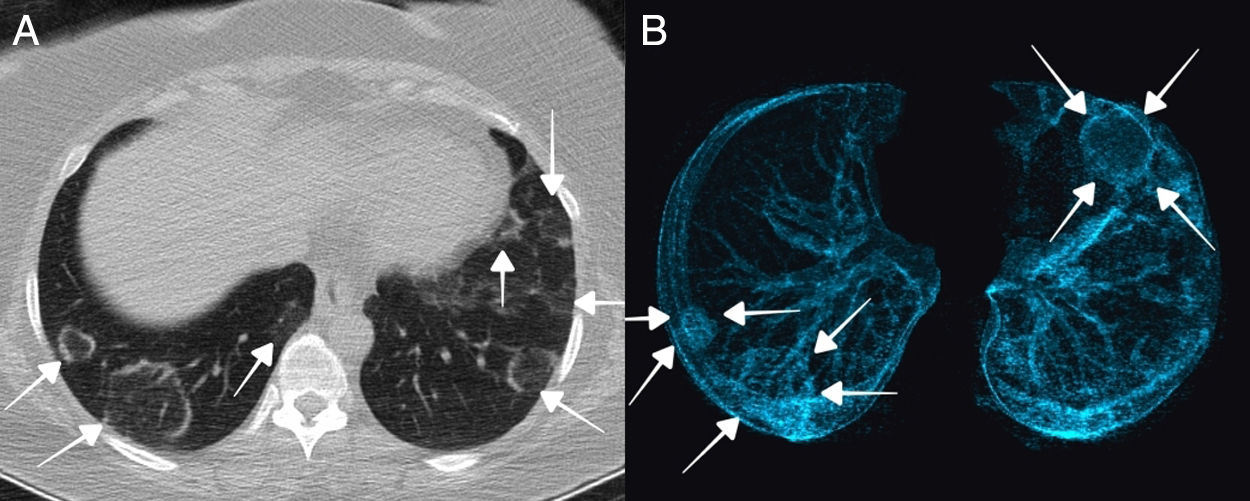
A 43-year-old female presented to the pandemic outpatient clinic of our hospital with the complaints of cough, diarrhea and loss of taste for 3 days. She is a healthcare worker and that her colleagues with whom she worked have been diagnosed with COVID-19. Vital signs and physical examination were normal. Laboratory tests revealed an increased D-Dimer level (1365ng/ml). Chest CT and 3D volume-rendering images demonstrated bilateral, multilobar peripherally placed lesions with a “multiple reversed halo sign” (Fig. 1A and B). A nasopharyngeal swab was positive for COVID-19. The patient was hospitalized and treated with hydroxychloroquine and enoxaparin. On day 7 of hospitalization, clinical symptoms improved and patient was discharged.
 and axial 3D volume-rendering (B) images revealed bilateral, multilobar peripherally located lesions with “multiple reversed halo sign” (arrows).")
The reversed halo sign is defined as an area with ground-glass opacity surrounded by partial or complete rings of consolidation.1 This sign is nonspecific and has been reported in cryptogenic, non-cryptogenic organizing pneumonia, viral and fungal pneumonia, neoplastic processes and many of other pathologies.2 This sign is often associated with severe cases and occurs long after symptom onset. Such findings in these cases suggest that non-neoplastic processes such as organized pneumonia are one of the mechanisms of lung fibrosis.2


