
Highly effective CFTR modulator therapy with elexacaftor–tezacaftor–ivacaftor (ETI) has revolutionized clinical outcomes in cystic fibrosis (CF), yet its effects on gut and lung microbiota, especially at the functional level, are poorly understood.
MethodsIn a 12-month prospective study, we enrolled 35 clinically stable CF patients initiating ETI. Paired fecal and sputum samples, collected at baseline and after 12 months, were analyzed using shotgun metagenomics, metaproteomics, and short-chain fatty acid (SCFA) quantification. Multi-omics data were integrated with clinical parameters assessing lung, hepatic, pancreatic, and intestinal function.
ResultsETI drove significant clinical improvements, including increased ppFEV1, higher fecal elastase, and better nutritional status, despite persistent major lung pathogens and minimal changes in liver or intestinal inflammation markers. Microbiota composition showed limited shifts: alpha diversity was stable, and beta diversity changes accounted for only small variance in both compartments. However, butyrate-producing genera enriched in feces, while oropharyngeal taxa increased in sputum. Metaproteomics revealed broad downregulation of host neutrophil-driven inflammatory proteins; sputum additionally showed increased abundance of extracellular matrix-related proteins. Microbial proteins linked to carbohydrate/lipid metabolism, particularly butanoate pathways, increased in feces alongside a trend for higher butyrate. In sputum, formaldehyde dehydrogenase enzymes rose, indicating enhanced oxidative microbial metabolism.
ConclusionsETI is associated with minimal compositional but substantial functional reprogramming in CF microbiota. These changes are accompanied by an increase in butyrate-producing taxa, attenuation of host pro-inflammatory pathways, and a shift in lung metabolism toward oxidation. Despite ongoing pathogenic colonization, these changes suggest CFTR modulation is associated with a less inflammatory, more stable host–microbiota ecosystem.
In recent years, CFTR modulators have changed the paradigm of cystic fibrosis (CF). The latest is a triple combination of two correctors (elexacaftor and tezacaftor) and a potentiator (ivacaftor) for people with at least one F508del mutation [1]. This treatment has led to an unprecedented improvement, significantly enhancing lung function, nutritional status, and quality of life compared to previous modulators [2]. However, its impact on the gut and lung microbiota, which in CF show reduced richness and diversity compared to healthy controls [3,4], remains understood, despite altered microbiota being linked to systemic inflammation. Most available microbiota studies have focused on composition, even though functionality is equally important. Addressing this requires integrating metagenomics, metaproteomics, and metabolomics to obtain a more comprehensive view of host–microbe interactions.
Our main hypothesis is that individual clinical changes (ppFEV1, AST/ALT/GGT, calprotectin, elastase) are associated with targeted changes in microbiological taxa and functions. Therefore, we first described compositional and functional profiles, and second, whether clinical improvements in lung, hepatic, intestinal, and pancreatic functions after one year of ETI are associated with specific bacterial changes.
Material and methodsStudy designA prospective descriptive study was conducted in our center between October 2021 and February 2024 (28 months). Thirty-five people with CF in a stable clinical phase were included after written informed consent and approval from the Ethics Committee of Ramón y Cajal Hospital (reference 328/2020). The cohort comprised individuals who had never received CFTR modulators and those previously treated before ETI initiation. Fecal and sputum samples were collected at baseline (T0), and after one year of treatment (T12) and stored at −80°C in separate aliquots of 0.5g (feces) or 1mL (sputum) until processing. Sputum was used for conventional cultures, and both sample types were used for metagenomic, metaproteomic, and metabolomics assays. Clinical characteristics, treatment data, including chronic inhaled antibiotics and azithromycin, and microbiological cultures were collected from medical records.
Metagenomic shotgun sequencingSample pre-processing, DNA extraction, library preparation and sequencing (Illumina NovaSeq-6000, 2×150bp), generation of demultiplexed FASTQ files, taxonomic profiling with MetaPhlAn4 v4.1.1, and downstream bioinformatic processing are described in Supplementary File 1.
MetaproteomicsFecal and sputum aliquots were processed following a modified protocol based on García-Durán et al. [5]. Full details of sample preprocessing and downstream bioinformatic analysis are provided in Supplementary File 1.
MetabolomicsFecal and sputum aliquots were processed as previously described [6]. Full details of sample preprocessing and targeted metabolomic analysis are provided in Supplementary File 1, with identification parameters listed in Supplementary Table S1A–B.
Statistical analysis and data availabilityFull details of the statistical analysis are provided in Supplementary File 1. Sequence data are available in the European Nucleotide Archive (BioProject PRJEB103799). Proteomics data are available via PRIDE (ProteomeXchange identifier PXD070696). All analysis code and data are freely available on GitHub (https://github.com/nbastonpaz/CF_ETI_multi-omics_gut_lung_microbiota) under a GPL-3.0 license.
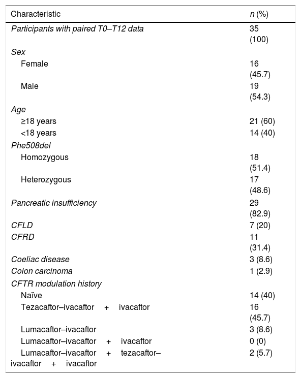
ResultsDescription of clinical characteristics and outcomesThirty-five individuals completed the 12-month follow-up. Baseline characteristics and clinical data are summarized in Table 1 and in Table S2. Lung function significantly improved at T12 (p<0.001, mean 70.3±SD 25.1 vs 81.5±23.7); half of patients (n=18) remained in the normal–mild category (>70% ppFEV1), 31.4% (n=11) in moderate–severe (<40–69% ppFEV1), and 17.1% (n=6) improved from moderate–severe to normal–mild. Lung function improved significantly from T0 to T12 within both sex subgroups [males (n=19): p<0.001, 73.4±24.7 vs 84.7±22.1; females (n=16): p=0.005, 66.6±25.8 vs 77.8±25.7], and age subgroups [adults (n=21): p<0.001, 57.2±19.1 vs 69.4±17.4; pediatrics (n=14): p=0.02, median 89.5 (85.5–97.8) vs 97.5 (88.8–109)] (Fig. S1). However, linear mixed-effects models showed no significant Time×Sex or Time×Age Group interaction, indicating that the magnitude of ppFEV1 improvement did not differ significantly between males and females or between adults and pediatric subjects.
Baseline demographic and genetic characteristics, reported as n (%).
| Characteristic | n (%) |
|---|---|
| Participants with paired T0–T12 data | 35 (100) |
| Sex | |
| Female | 16 (45.7) |
| Male | 19 (54.3) |
| Age | |
| ≥18 years | 21 (60) |
| <18 years | 14 (40) |
| Phe508del | |
| Homozygous | 18 (51.4) |
| Heterozygous | 17 (48.6) |
| Pancreatic insufficiency | 29 (82.9) |
| CFLD | 7 (20) |
| CFRD | 11 (31.4) |
| Coeliac disease | 3 (8.6) |
| Colon carcinoma | 1 (2.9) |
| CFTR modulation history | |
| Naïve | 14 (40) |
| Tezacaftor–ivacaftor+ivacaftor | 16 (45.7) |
| Lumacaftor–ivacaftor | 3 (8.6) |
| Lumacaftor–ivacaftor+ivacaftor | 0 (0) |
| Lumacaftor–ivacaftor+tezacaftor–ivacaftor+ivacaftor | 2 (5.7) |
Abbreviations: CFLD, cystic fibrosis-related liver disease; CFRD, cystic fibrosis-related diabetes.
Within-patient changes in different clinical measures over 12 months are shown in Table S3. Transaminase and fecal calprotectin levels did not change significantly, whereas fecal elastase increased significantly. BMI significantly improved in adults, while SD z-score increased in children, but not significantly (Fig. S2).
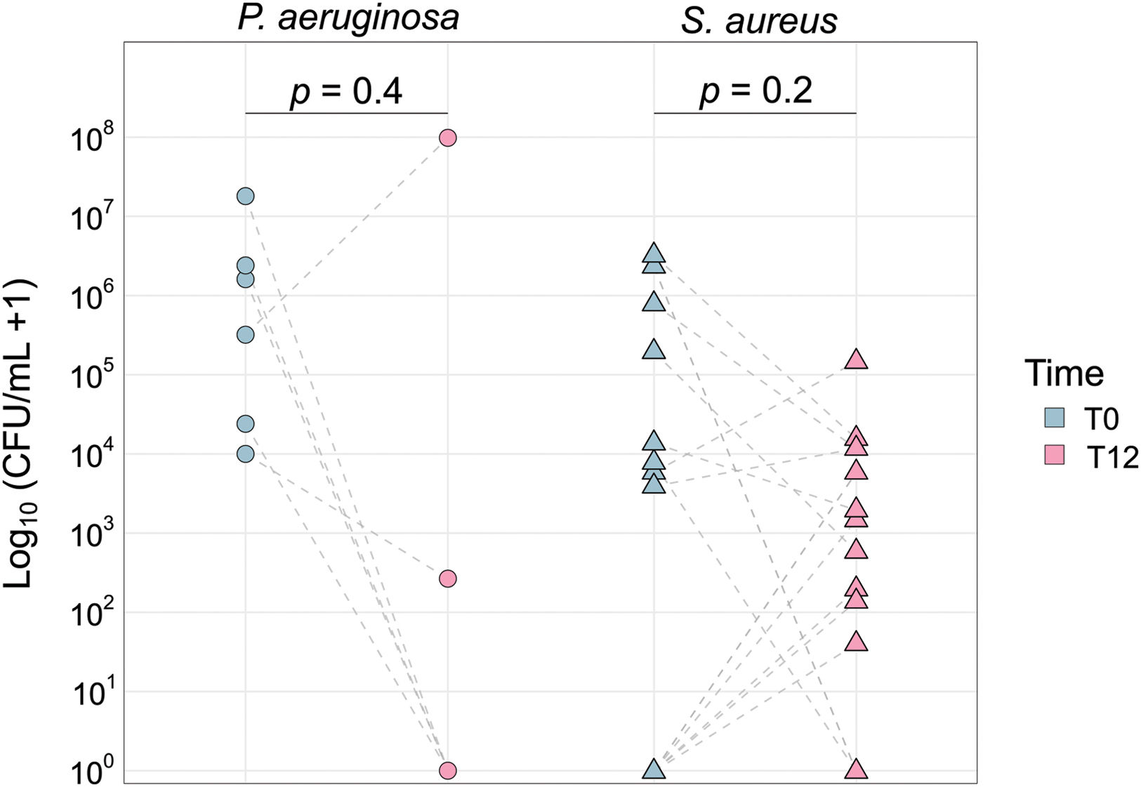
Overall, the density of Pseudomonas aeruginosa colonization decreased, and Staphylococcus aureus increased after treatment, although neither change was statistically significant (Fig. 1). Most patients with increased S. aureus (6/8) also improved lung function, but the proportion of patients with improved ppFEV1 was similar regardless of S. aureus increase (Fisher's exact test, p=1.0). The eight patients co-colonized by P. aeruginosa and S. aureus showed individualized trajectories (Fig. S3).
Composition and functional microbiome profiles
One fecal and five sputum samples were excluded due to failure of library generation. To maintain paired analysis, timepoint samples from one patient (P3) were removed from the fecal dataset, and from five patients (A12, A20, A26, P16, and P21) were removed from the sputum dataset. This pairing was preserved across metaproteomic, metabolomic, and clinical models’ analysis. For the final metagenomic dataset, sequencing depth and read retention after quality filtering and host read removal are summarized in Table S4.
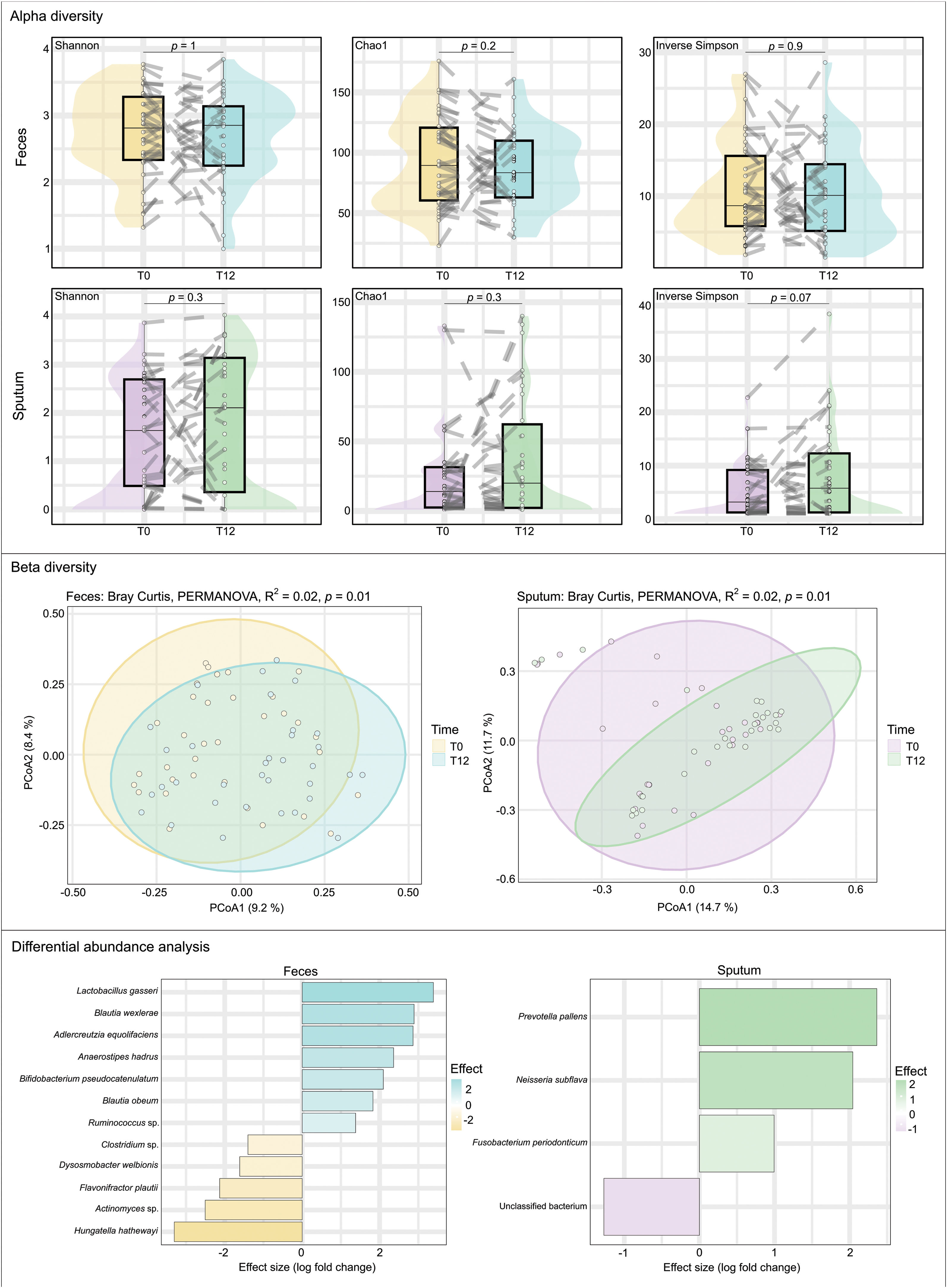
MetagenomicsNo significant differences in alpha diversity indices were detected in feces or sputum (Fig. 2). In contrast, differences in beta diversity (Bray–Curtis) were significant (feces: p=0.006; sputum: p=0.008), although explained variance was low (R2=0.025 and R2=0.023, respectively) (Fig. 2). The composition of the bacterial community was largely similar between timepoints, with modest shifts in specific taxa (Fig. 2). For instance, in feces, Blautia, Adlercreutzia, Anaerostipes, Bifidobacterium, and Ruminococcus were more abundant at T12. The mean relative abundance of Blautia wexlerae, Bifidobacterium pseudocatenulatum and Anaerostipes hadrus increased from 1.8%, 1.6%, and 1.7% at T0 to 5.5%, 2.1%, and 3.9% at T12, respectively (Table S5). Conversely, Adlercreutzia equolifaciens and Ruminococcus sp. had low prevalence and mean abundances (<0.2% at both timepoints). In sputum, typical oral taxa from genera Prevotella, Neisseria, and Fusobacterium were more abundant at T12. Overall, the community composition remained broadly similar between timepoints in both compartments, with only modest shifts in specific taxa.
Metaproteomics and targeted metabolomics
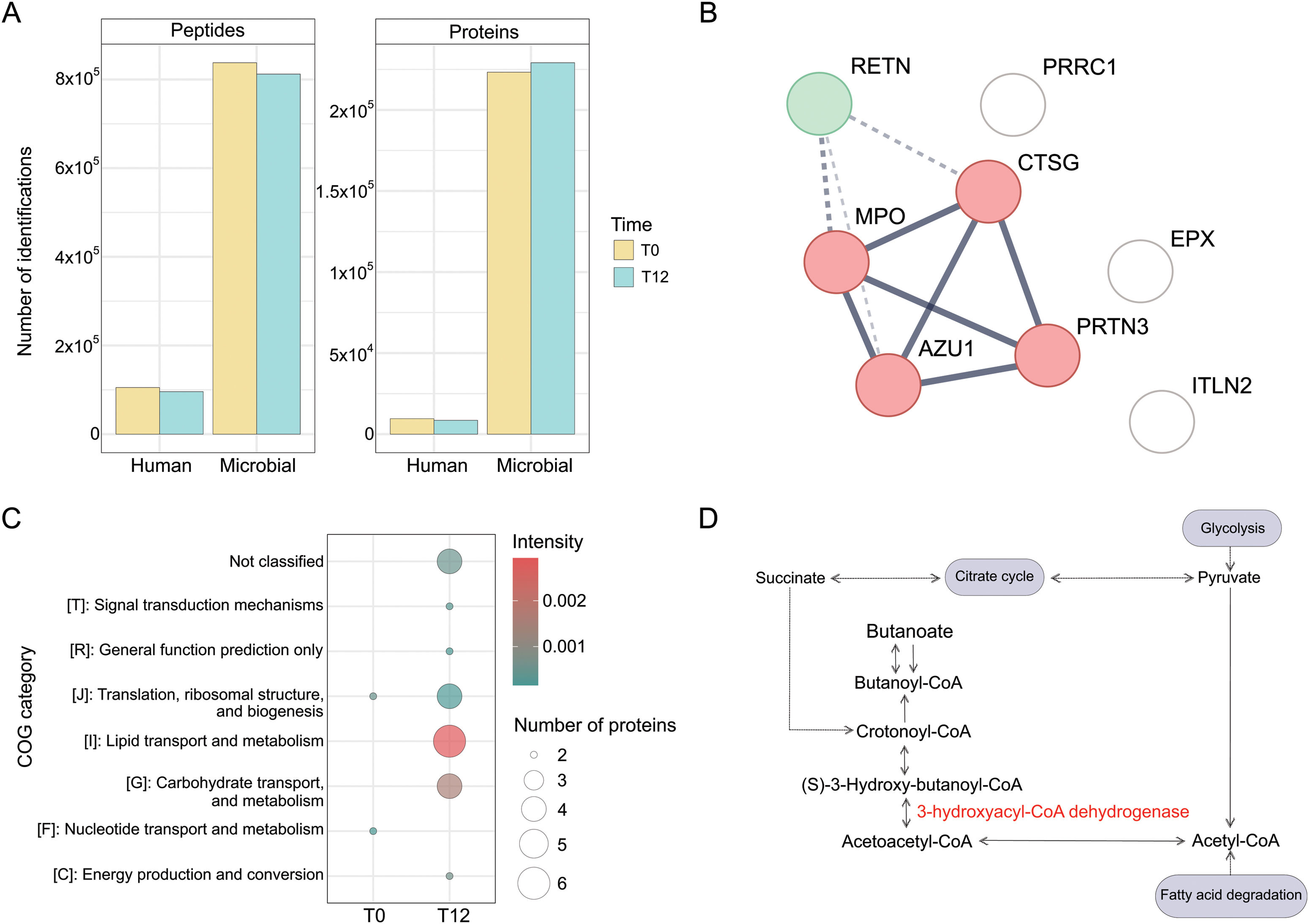
In feces, human inflammation-associated proteins were significantly enriched at T0. Clustering identified two groups: neutrophil-associated proteins and resistin (RETN), a proinflammatory cytokine (Fig. 3B). Calprotectin tended to decrease (LFC=−0.96, p-adj=0.056). At T12 intelectin-2 (ITLN-2), involved in cellular recognition and defense against pathogens, increased significantly (LFC=0.99, p-adj=0.03).

Metaproteomic fecal analysis. (A) Account for the number of human and microbial proteins and peptides. (B) Full STRING protein–protein interaction network of significant human fecal proteins grouped into two clusters. Cluster 1 (red): neutrophil-associated proteins. Cluster 2 (green): resistin protein (RETN). Each node represents all the proteins produced by a single, protein-coding gene locus. Edges represent protein–protein interactions that jointly contribute to a shared function. Line thickness indicates the strength of data support. Dotted lines indicate the edges between clusters. (C) COG functional categories of microbial fecal proteins at T0 and T12. Dot size indicates the number of proteins, and color represents average intensity. (D) Summary diagram of butyrate metabolism, highlighting the role of the enzyme 3-hydroxyacyl-CoA dehydrogenase. Detailed results are shown in Table S6.
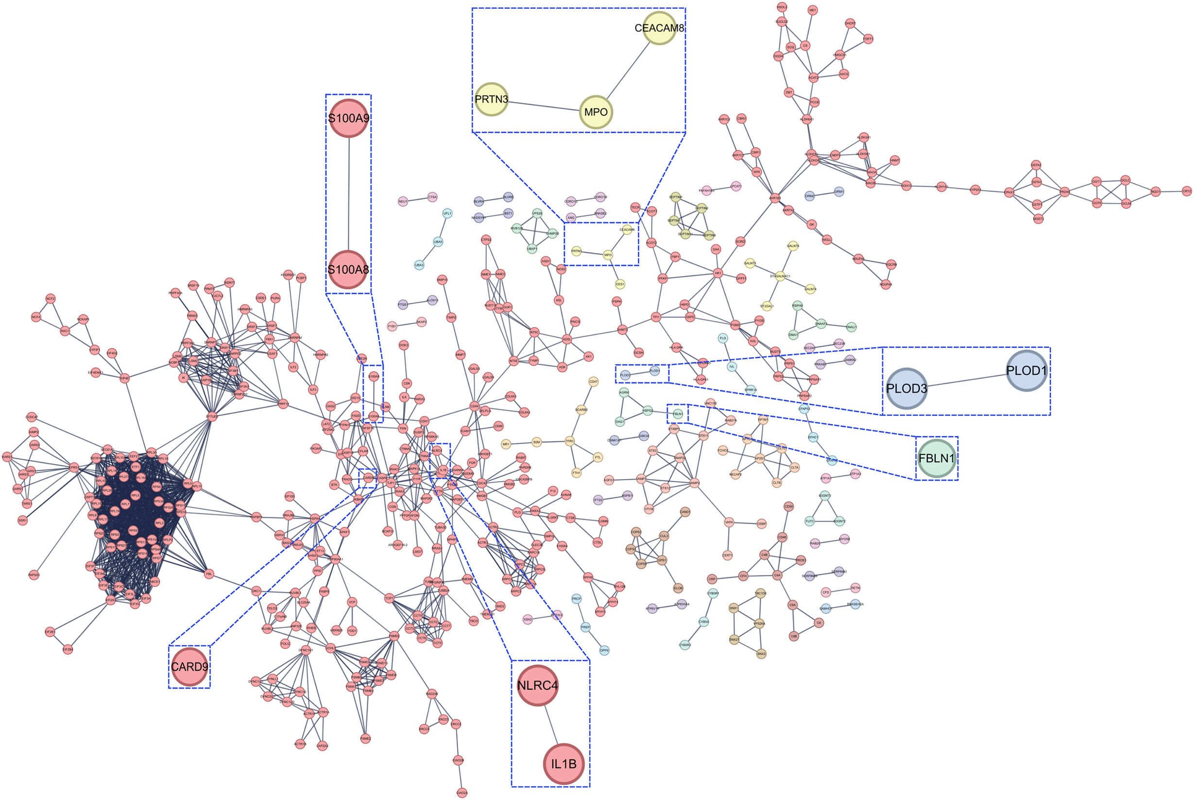
In sputum, abundance changes in 914 human proteins were significant (Fig. 4). Proteins associated with innate immunity and chronic inflammation decreased, particularly neutrophil markers: calprotectin subunits S100A8 and S100A9 (LFC=−0.66, p-adj=0.003; LFC=−0.70, p-adj=0.01), myeloperoxidase (MPO; LFC=−0.68, p-adj=0.01), proteinase 3 (PRTN3; LFC=−0.54, p-adj=0.02) and the neutrophil adhesion molecule CEACAM8 (LFC=−0.96, p-adj=0.01). The pro-inflammatory cytokine interleukin-1 beta (IL1B) was also strongly downregulated (LFC=−1.63, p-adj=0.02). Innate immune signaling proteins NLRC4 (LFC=−1.16, p-adj=0.008) and CARD9 (LFC=−2.68, p-adj=0.02), were also significantly reduced. Conversely, extracellular matrix-related proteins were significantly more abundant at T12, such as procollagen-lysine 5-dioxygenase 1 (PLOD1) and 3 (PLOD3) (LFC=0.74, p-adj=0.02; and LFC=2.03, p-adj=0.04, respectively), and fibulin-1 (FBLN1, LFC=1.44, p-adj=0.01).

Full STRING protein–protein interaction network of significant human sputum proteins grouped into 40 clusters, being the greatest cluster including proteins associated with innate immunity and protein homeostasis. Each node represents all the proteins produced by a single, protein-coding gene locus. Edges represent protein–protein associations, jointly contributing to a shared function. Line thickness indicates the strength of data support. Dotted lines indicate the edges between clusters. Highlighted areas (zoomed boxes) correspond to proteins related to inflammation and fibrosis. Detailed results are shown in Table S7.
In feces, few microbial proteins were detected at T0, whereas at T12 both diversity (number of proteins) and abundance (intensity) associated with carbohydrate transport/metabolism (COG G) and lipid transport/metabolism (COG I) increased, notably 3-hydroxyacyl-CoA dehydrogenase, implicated in butyrate metabolism (LFC=0.81, p-adj=0.03) (Fig. 3C and D). Fecal butyrate showed a non-significant trend toward higher levels, opposite to sputum, where butyrate tended to decrease (Fig. S4).
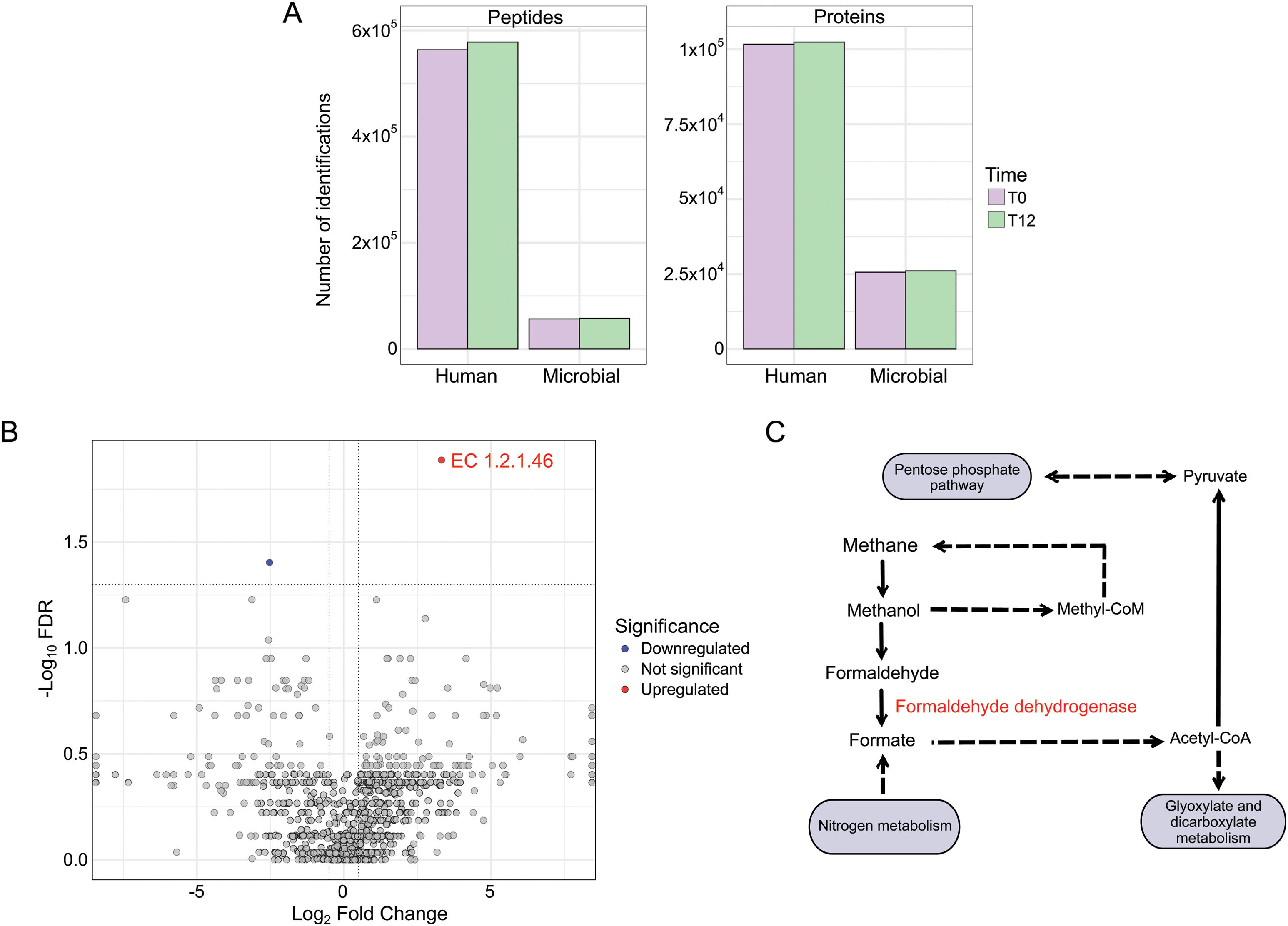
In sputum, no individual microbial protein reached significance. However, grouping by enzyme activity revealed significant changes: enzymes annotated as formaldehyde dehydrogenase (EC 1.2.1.46), an oxidoreductase that catalyzes the oxidation of formaldehyde to formate and participates in methane metabolism, were more abundant at T12 (LFC=3.32, p-adj=0.01) (Fig. 5B).

Metaproteomic sputum analysis (Table S7). (A) Account for the number of human and microbial proteins and peptides. (B) Volcano plot of differentially abundant microbial protein functional categories in sputum metaproteomes after one year of ETI therapy. The x-axis displays the log2 fold change, and the y-axis the −log10 FDR. Each point represents a microbial enzyme classified by its EC number. (C) Summary diagram of methane metabolism, highlighting the role of the enzyme formaldehyde dehydrogenase.
All significant associations are reported in Table S8. In feces (Fig. S5), Akkermansia muciniphila and Bifidobacterium pseudocatenulatum were positively associated with better lung function before treatment (both LFC=0.18, p-adj=0.04). Veillonella atypica and Veillonella rogosae were negatively associated with elastase at baseline (LFC=−1.38 and −6.63, both p-adj<0.001). No significant associations were found between specific taxa and calprotectin. Across transaminase, Clostridium species were negatively associated with ALT and AST after treatment (p-adj=0.03 and p-adj<0.001, respectively), whereas Clostridium symbiosum was positively associated with GGT both before and after treatment (LFC=0.02 at T0 and 0.04 at T12, both p-adj<0.001).
In sputum (Fig. S6), no significant association between taxa and lung function was observed before or after treatment, but several relationships with other clinical variables were detected. Before treatment, S. aureus was more abundant in patients with lower elastase and calprotectin (LFC=−0.26, p-adj<0.001; LFC=−0.04, p-adj=0.04) and showed positive associations with AST and GGT (LFC=0.24, p-adj=0.03; LFC=0.16, p-adj<0.001).
After 12 months, Fusobacterium species (F. nucleatum and F. periodonticum) were positively associated with ALT and AST (LFC=0.14 and 0.02, both p-adj<0.001), while F. nucleatum showed a strong negative association with GGT at baseline (LFC=−0.62, p-adj<0.001).
Within genus Veillonella, patterns differed by species and transaminase: V. parvula and V. tobetsuensis were positively associated with AST at T12 (LFC=0.07 and 0.15, both p-adj=0.01), whereas V. atypica was negatively associated (LFC=−0.08, p-adj=0.002). For GGT, V. atypica showed a negative association both before and after treatment (LFC=−0.03 at T0 and −0.22 at T12, both p-adj<0.001), similar to V. dispar (LFC=−0.09 at T0 and −0.21 at T12, both p-adj<0.001).
Prevotella species also showed opposing patterns. Prevotella histicola and P. nanceiensis were negatively associated with AST at T12, whereas P. salivae showed a positive association (LFC=−0.28, p-adj=0.004; LFC=−0.05, p-adj=0.01; LFC=0.05, p-adj=0.04). Prevotella salivae and P. pallens were negatively associated with GGT at T12 (LFC=−0.22, p-adj<0.001; LFC=−0.07, p-adj=0.02), in contrast to P. melaninogenica, which was positively associated with GGT after treatment (LFC=0.04, p-adj<0.001).
DiscussionCFTR modulators have transformed CF management since ETI became available [7]. In this study, we assessed the impact of one year of ETI on gut and lung microbial communities and whether individual clinical improvements were associated with specific bacterial changes. ETI led to clear clinical improvement, particularly in lung function [8], with almost 20% of our patients shifting from moderate–severe to normal–mild stage. Gastrointestinal improvement was more modest, although we also observed signs compatible with recovery from pancreatic insufficiency to sufficiency in some patients [9,10]. Fecal elastase levels increased significantly, whereas calprotectin did not change. The absence of a significant change in fecal calprotectin may be explained by the clinically stable status of the cohort at baseline and by the fact that paired calprotectin values were not available for all patients, reducing statistical power for this analysis. Both adults (significantly) and children (trend) improved BMI, as previously reported [11,12]. Transaminases did not change significantly, although AST showed a tendency to increase, possibly attributed to therapy [13].
Eradication of the main CF-colonizing pathogens was also not observed after 12 months of ETI [14,15]. In our cohort, the P. aeruginosa load tended to decline, whereas S. aureus load tended to increase. Most patients whose S. aureus load increased tended to improve lung function. In mixed colonization, the reduction of P. aeruginosa may diminish ecological competition, facilitating the growth of S. aureus. Evidence suggests that S. aureus may play different roles depending on age and disease stage in CF: it has been associated with airway inflammation and reduced lung function in children [16], but in adults may indicate a less severe state, particularly when P. aeruginosa colonization decreases. Eradication of non-tuberculous Mycobacteria has been reported in a multicenter study [17], including M. abscessus. We observed this in two patients; however, another remained colonized. Although the main pathogens were not eliminated, other atypical microorganisms may increase. Despite ETI-related improvements in mucociliary clearance, residual bronchial structural damage may still impair airway defenses and facilitate colonization by upper respiratory tract microorganisms.
Despite the clinical improvements, overall gut microbiota diversity remained relatively stable. Alpha diversity did not change significantly, and beta-diversity shifts explained only a small fraction of variance. However, these findings reflect the 12-month post-treatment state and do not exclude earlier transient microbial changes after ETI initiation. Similar findings were reported by Gutiérrez-Díaz et al. [18], whereas Reasoner et al. [19] described increased alpha diversity in patients aged 6–18 years. We observed specific compositional changes, especially in feces, where enrichment of the butyrate-producing bacterium Anaerostipes hadrus was significant after one year of treatment. Butyrate-producing bacteria have been linked to reduced gut inflammation [20], and SCFAs, particularly butyrate, have beneficial local and systemic effects, especially on the respiratory tract [21,22]. These findings suggest that ETI may promote an increase in beneficial taxa. In sputum, compositional changes were more discrete and mainly involved commensal oropharyngeal species. The high proportion of host reads in sputum samples reflects the difficulty of obtaining high-quality sputum samples after ETI, when sputum production may decrease. This may also be consistent with the increased detection of oropharyngeal-associated taxa at T12. Previous studies have reported ETI-induced shifts in the respiratory microbiota, with increased evenness, patient-specific community signatures, and early increases in commensal taxa shortly after treatment initiation [23,24].
Functional metaproteomic analyses revealed more distinct differences than compositional data. After 12 months of ETI, the intestinal and airway environments appeared to shift from a pro-inflammatory to a more balanced state. In fecal samples, neutrophil-associated human proteins and RETN, a myeloid-derived proinflammatory cytokine [25], were significantly more abundant at baseline. After treatment, ITLN2 was the only human protein significantly more abundant; this Paneth cell–specific protein, reduced in inflammatory bowel disease [26], is consistent with a more regulated mucosal environment. In sputum, we observed a global downregulation of inflammatory and innate immune proteins after ETI. By contrast, extracellular matrix-associated proteins remained upregulated, which may be compatible with residual airway structural remodeling in the context of chronic CF lung disease. This structural remodeling may contribute to the persistence of pathogenic populations and the immigration of oropharyngeal organisms. Consistently, systemic inflammation is reduced with ETI, but the immune profile of these patients remains different from that of healthy controls [27].
The fecal microbial metaproteome increased in proteins associated with carbohydrate and lipid metabolism. Although fecal butyrate did not increase significantly, enzymes in butanoate metabolism, particularly 3-hydroxyacyl-CoA dehydrogenase (EC 1.1.1.35), were significantly elevated. These findings are consistent with the enrichment of butyrate-producing bacteria and indicate that a stable microbial community can shift its functional output depending on the surrounding physiological context. In sputum, no individual microbial protein reached significance, but grouping by enzyme activity revealed a significant increase in formaldehyde dehydrogenase (EC 1.2.1.46). Non-methylotrophic bacteria such as P. aeruginosa can oxidize formaldehyde directly to formate via glutathione-independent pathways [28], and the upregulation of this enzyme may reflect a shift toward more oxidative microbial metabolism, possibly driven by reduced mucus obstruction and better oxygen availability after treatment.
Several fecal taxa were associated with clinical status. Before treatment, A. muciniphila and B. pseudocatenulatum were enriched in individuals with better lung function, and these differences disappeared after one year of ETI. Regarding pancreatic function, V. atypica and V. rogosae were more abundant in individuals with lower elastase levels at baseline; V. atypica remained enriched in this group after 12 months. Although this genus has not been directly related to pancreatic insufficiency, it has been reported as enriched in the fecal microbiome of pancreatic cancer patients [29]. Transaminase levels did not change significantly overall, but associations with fecal taxa were species-specific: several Clostridium species were more abundant in individuals with lower ALT and AST after treatment, whereas C. symbiosum was enriched in patients with higher GGT.
In sputum, no specific taxa were correlated with improved lung function before or after treatment, which is expected, as the clinical effects are likely driven by community-wide rather than single-species changes. However, S. aureus was more enriched in patients with lower elastase and calprotectin and with higher AST and GGT at baseline, although all these associations disappeared after one year, despite an overall increase in S. aureus counts. A less inflamed airway environment and reduced competition with P. aeruginosa may allow S. aureus to persist without necessarily indicating worse clinical status. As in feces, associations between microbial taxa and transaminases were species-specific. Fusobacterium spp. were more abundant in patients with higher ALT and AST after treatment, but F. nucleatum was enriched in those with lower GGT. Similar genus- and species-specific patterns were observed for Veillonella and Prevotella. These context-dependent relationships suggest that anaerobes in CF act as adaptable members of a dynamic microbial network rather than being strictly beneficial or harmful. Overall, these associations should be considered exploratory and intended to identify potential clinical–microbiome relationships rather than causal effects.
As limitations, the unadjusted T0–T12 compositional and functional analyses are descriptive and do not identify adjusted causal effects. These analyses were intended to capture broad patterns over one year of ETI, rather than age-stratified responses, which were not assessed because further subgrouping would have reduced statistical power in this moderate-sized longitudinal cohort. Clinical–microbiome association models were then used to examine individual clinical variables, with age and sex included as covariates. However, we did not adjust for prior CFTR modulator therapy or CFTR genotype, as Reasoner et al. [19] reported similar community composition in previously treated and modulator-naïve individuals, and Duong et al. [30] found genotype-related differences explaining only ∼2% of the variance. Similarly, antibiotic exposure was not included as an adjustment variable because treatment patterns changed during follow-up and were highly unbalanced at T12, particularly for azithromycin. Such additional adjustment or stratification in our cohort would have further reduced statistical power due to our limited sample size. Moreover, some differentially abundant taxa, particularly in feces, were of low relative abundance and should be interpreted cautiously. Our cohort consisted of clinically stable patients at baseline, which may have limited the magnitude of microbiota or inflammatory changes observed. Other systemic inflammatory markers, such as CRP, which may decrease after ETI initiation, and cardiometabolic risk variables were not incorporated because they were not systematically available at paired T0 and T12 timepoints. Additionally, sputum quality, especially at 12 months, may have been suboptimal and more representative of upper than lower respiratory airway microbiota. Finally, the absence of intermediate paired fecal and sputum multi-omics samples prevented the assessment of early transient microbial shifts, including possible short-term changes in alpha- or beta-diversity that may have stabilized before T12. A longer follow-up is also needed to determine whether the observed changes are sustained over time.
In summary, this longitudinal multi-omics study shows that one year of ETI therapy produces substantial clinical improvement in people with CF, accompanied by targeted microbiome and metaproteome functional shifts rather than global community restructuring. Specific gut taxonomic and functional adaptations appear linked to butyrate-producing bacteria, butyrate-related pathways, and downregulation of inflammatory human proteins in both compartments. Following respiratory improvement, oxidative metabolism gains prevalence over fermentation in the lungs. Species-specific roles of anaerobic genera must be considered, although further studies are needed to determine causality. Despite persistent pathogenic colonization, probably due to permanent fibrotic damage, our results support that CFTR modulation delivers its systemic benefits not by resetting the microbiota but by functionally reprogramming the host–microbe ecosystem toward a less harmful state.
Authors’ contributionsN.B.P. conceptualization and writing original draft; N.B.P., C.G.D., E.P.G., A.M.B. experimental processing and writing; N.B.P., D.B., and J.T. metagenomic analysis; N.B.P. and C.G.D. metaproteomic analysis; J.T. and A.C.S. supervision in metagenomic analysis, writing – review, and editing; M.G., L.M., and S.V.S. providing resources and validation; A.O., J.O.I., R.C. and C.G. writing – review and editing; J.D.D.C., M.G., and R.d.C. conceptualization, writing – review and editing; A.O., J.O.I., R.d.C., S.V.S., and J.D.D.C. funding acquisition.
Ethical committeeThe Hospital Ramón y Cajal Ethics Committee approved in 2020 (Ref. number 328/2020). The research was conducted ethically, in accordance with the World Medical Association Declaration of Helsinki. All participants and their parents/legal guardians signed a written informed consent form.
Declaration of generative AI and AI-assisted technologies in the writing processArtificial intelligence has not been used in the design of the article or in the creation of images, but the manuscript has been grammatically and orthographically corrected using the deepL website (https://www.deepl.com/es/translator).
FundingThis work was supported by the Instituto de Salud Carlos III and the European Union – NextGenerationEU (PI22/01045 to J.D.D.C. and S.V.S., and PI19/01043 to R.C. N.B.P. was supported by a research contract from Instituto de Salud Carlos III (Rio Hortega program, grant number CM23/00037). This research was also supported by Personalized and precision medicine grant from the Instituto de Salud Carlos III (MePRAM Project, PMP22/00092), Instituto de Salud Carlos III, Ministerio de Ciencia e Innovación, funded by NextGenerationEU funds from the European Union that finance the actions of The Resilience and Recovery Facility.
Declaration of competing interestL.M., S.V.S., and R.d.C. have received funding from Vertex S.L. The rest of the authors declare that the research was conducted in the absence of any commercial or financial relationships that could be construed as a potential conflict of interest.
We thank Dr Leticia Olavarrieta Scappini and the technical staff of UCA-GT for their assistance with the library's construction and metagenomic sequencing. We deeply thank Raquel Barbero-Herránz and Dr Val Fernández from IRYCIS for their valuable advice in metagenomic and clinical correlation analysis. We also thank Dr Felipe Clemente, Inés Zapico, and Samuel de la Cámara for their insightful help with the LC-MS/MS analysis and data curation. We sincerely thank Dr Fernando Baquero Mochales for his valuable suggestions and corrections. Lastly, we acknowledge the people with CF and the families of the children for their participation, and all the health personnel of the CF unit of Ramón y Cajal University Hospital.
The following are the supplementary data to this article: