

Electrical impedance tomography (EIT) is a radiation-free, non-invasive, real-time bedside technique that maps the regional distribution of lung ventilation. By tracking the small changes in electrical impedance that accompany the movement of air, it produces a dynamic, regional picture of where ventilation goes and surrogately, how ventilation and perfusion into the lungs are distributed [1].
It came of age largely as a promising monitoring tool in intensive care units (ICU), used most frequently for individualized positive end-expiratory pressure (PEEP) titration and recruitment assessment. In mechanically ventilated ARDS patients with coexisting COPD, EIT-guided PEEP titration selected pressures a third lower than ARDSnet (6.9±1.7 vs. 10.4±1.2cmH2O), with a lower mechanical power (13.9 vs. 15.9J/min) and global inhomogeneity index (GI: 0.40±0.04 vs. 0.45±0.05, p=0.001); far from the haemodynamic penalty a lower PEEP might be expected to carry in obstructive disease, oxygen delivery was greater [2]. However, its most compelling potential may lie elsewhere: in becoming the main tool of pneumologists and physiotherapists at bedside, turning some invisible processes into something we can see, measure and adjust in the moment. This would be a shift from EIT as a passive monitor to EIT as an active guide for individualized interventions; in short, from monitoring to precision respiratory support.
For decades we have titrated non-invasive ventilation (NIV), applied airway clearance techniques and mechanical insufflation–exsufflation (MI-E) on global surrogates such as tidal volume or oxygen saturation and, commonly for physiotherapists, auscultation. Too often, on a fixed protocol applied uniformly to different lungs and patients. However, a pressure that recruits a dependent dorsal region may over-distend an already hyperinflated apex; a compression that drains one lobe may impair ventilation in another; an insufflation–exsufflation pressure setting that mobilises secretions may simultaneously provoke dynamic airway collapse or overdistension. When we cannot see the regional consequences of interventions, we cannot know whether we are helping one part of the lung at the expense of another.
That regional blindness is especially consequential in COPD. The EIT-derived GI seems already higher in subjects with preserved ratio impaired spirometry than in those with normal spirometry, with COPD values overlapping both [3]. Measured during the spirometry, GI of FEV1 and of FEV1/FVC correlated with the change in CAT score (R=0.62, p=0.002 and R=0.68, p<0.001), while the corresponding changes in spirometry did not (FEV1 R=0.1; FEV1/FVC R=−0.01) [4]. If a global forced vital capacity can miss what the patient reports, so can a global volume measurement; EIT restores the regional view that bedside needs.
Evidence for EIT-guided NIV titration is now beginning to appear. In intubated COPD patients, regional expiratory time constants estimated by EIT vary from 2 to 5s across the lung, and a 2cmH2O PEEP step may improve one region while worsening another in the same patient [5]; while these results come from ICU, the same physiology governs expiratory pressure set during NIV, which is chosen to make emptying more homogeneous and to limit pendelluft, the intra-tidal redistribution of gas between fast and slow compartments, and not only to recruit.
What EIT adds is to confirm whether a modest rise in expiratory pressure improves regional ventilation and reduces heterogeneity without sacrificing tidal volume or comfort, rather than inferring it from the ventilator screen alone. Two small, randomized studies bear this out. In 13 sedated children, NIV shifted ventilation to ventral regions increasing the center of ventilation (CoV) by 2.7% (95%CI: 0.5–4.5; p=0.017) compared to spontaneous breathing and by 2.5% compared to CPAP (95%CI: 0.01–5.5; p=0.043) and restored homogeneity by lowering the GI (p=0.035) and restoring end expiratory lung volume (EELV; p=0.002) [6]; these gains were lost once NIV was withdrawn, maybe explained because support was only applied 20min. Likewise, in 16 patients after cardiac surgery, NIV produced greater pulmonary aeration than oxygen therapy (p<0.05) and nearly twice the swing in GI (7.96 vs. 4.17, p=0.03) [7]. The same applies to high flow therapy (HFT), where the pressure reaching the lung is neither set nor displayed: HFT raised EELV and homogenised regional ventilation [8], adding 169mL [93–656] after extubation, with no change in the dorsal predominance of ventilation [9]. Where that volume goes, however, is not predictable. In 15 patients with stabilized hypercapnic exacerbation, most of them COPD, NIV increased EELV 443±426mL against 53±288mL with HFT at 30L/min (p=0.004) and 139±327mL at 50L/min (p=0.024) but the ventral–dorsal difference in ventilation remained similar throughout interventions (p=0.46) [10].
The literature on EIT-guided physiotherapy remains sparse, and most of it has developed indirectly in the ICU, as a by-product of EIT's primary role there. EIT has already captured the regional effects of early mobilisation, which shifts ventilation toward dorsal regions [11], and a small but growing literature now examines its value for assessing physiotherapy techniques in critically ill and difficult-to-wean patients [12–14]. In airway clearance, and in manual chest physiotherapy in particular, EIT may identify the hypoventilated region before the physiotherapist begins, inform the choice of technique and “dose”, and then confirm immediately whether ventilation in the targeted zone and GI have improved. During MI-E, perhaps the most promising application, EIT can verify that insufflation pressures recruit collapsed dorsal regions while exsufflation pressures clear secretions without provoking derecruitment: in six children with spinal muscular atrophy, lung volume during tidal breathing increased after MI-E in four of them (58.8±55.1mL) [15], allowing to walk the narrow line between effective and harmful and to individualise settings instead of one-size-fit-all pressures.
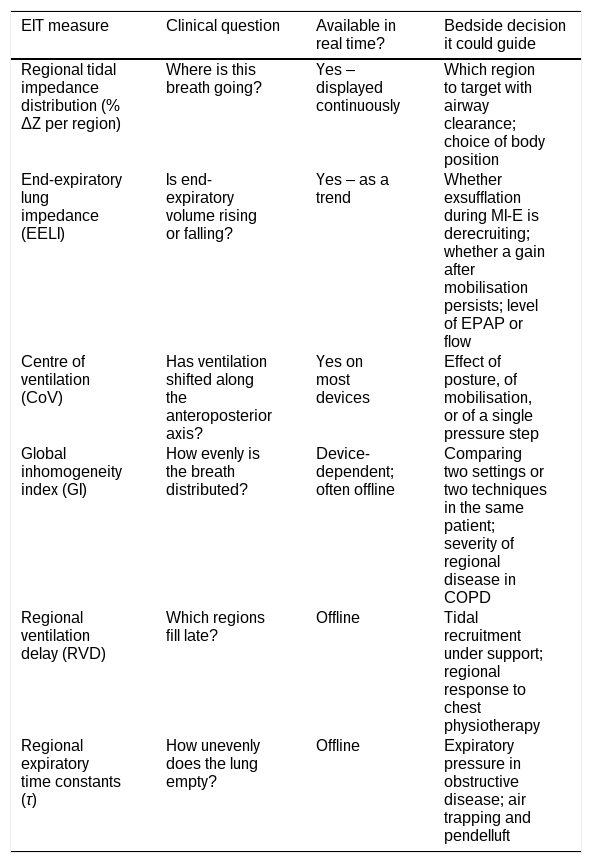
Whether these regional changes translate into outcomes that matter to patients remains unproven. This transition from monitoring to precision respiratory support needs clinical trials that test whether EIT-guided interventions improve clinical outcomes (e.g. secretion clearance, dyspnoea, length of stay) and not merely whether they shift an impedance curve. We also need consensus on which EIT-derived metrics matter, how to standardize them, and which patients stand to benefit most (Table 1). The promise of EIT is not the technology but the possibility of giving each patient the right intervention, measured and adaptive, at the right intensity.
Electrical impedance tomography measures and its availability at the bedside in real time or requires post-processing. Availability is device-dependent and should be checked against the specific system in use.
| EIT measure | Clinical question | Available in real time? | Bedside decision it could guide |
|---|---|---|---|
| Regional tidal impedance distribution (% ΔZ per region) | Where is this breath going? | Yes – displayed continuously | Which region to target with airway clearance; choice of body position |
| End-expiratory lung impedance (EELI) | Is end-expiratory volume rising or falling? | Yes – as a trend | Whether exsufflation during MI-E is derecruiting; whether a gain after mobilisation persists; level of EPAP or flow |
| Centre of ventilation (CoV) | Has ventilation shifted along the anteroposterior axis? | Yes on most devices | Effect of posture, of mobilisation, or of a single pressure step |
| Global inhomogeneity index (GI) | How evenly is the breath distributed? | Device-dependent; often offline | Comparing two settings or two techniques in the same patient; severity of regional disease in COPD |
| Regional ventilation delay (RVD) | Which regions fill late? | Offline | Tidal recruitment under support; regional response to chest physiotherapy |
| Regional expiratory time constants (τ) | How unevenly does the lung empty? | Offline | Expiratory pressure in obstructive disease; air trapping and pendelluft |
Artificial intelligence tools were used solely to rephrase selected sentences for readability, without altering the message, content or nature of the article. Authors reviewed and approved the final text and take full responsibility for it.
FundingThis research did not receive any specific grant from funding agencies in the public, commercial, or not-for-profit sectors.
Conflicts of interestThe authors declare not to have any conflicts of interest that may be considered to influence directly or indirectly the content of the manuscript.