


We report the case of a 66-year-old man with a history of bronchiectasis since his youth due to bronchopneumonia in childhood, untreated to date. He has self-limiting episodes of recurrent hemoptysis that are treated with antibiotics. He was admitted for a 20-day history of non-threatening hemoptysis.
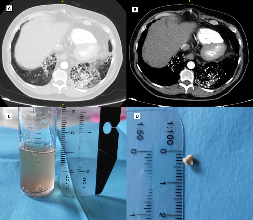
Computed tomography (CT): multiple bronchiectasis in lower lobes, with areas of atelectasis and broncholithiasis (Fig. 1A and B). FACED score: 5 points.
 Computed tomography with lung parenchymal window, axial slice. (B) Computed tomography with mediastinal window, axial slice. Cylindrical bronchiectasis predominantly in both lung bases, containing multiple calcified hyperdense images consistent with broncholiths. (C) A calcified, rounded body with slightly irregular edges measuring 4×3mm consistent with broncholith found in the bronchial aspiration tube. (D) Same fresh specimen.")
(A) Computed tomography with lung parenchymal window, axial slice. (B) Computed tomography with mediastinal window, axial slice. Cylindrical bronchiectasis predominantly in both lung bases, containing multiple calcified hyperdense images consistent with broncholiths. (C) A calcified, rounded body with slightly irregular edges measuring 4×3mm consistent with broncholith found in the bronchial aspiration tube. (D) Same fresh specimen.
In the targeted medical history, the patient reported that he has had recurrent episodes of expectoration of whitish, foul-smelling stones (lithoptysis) for years. During fiberoptic bronchoscopy, an irregular broncholith of 4×3mm was retrieved (Fig. 1C and D).
Broncholithiasis is a very rare disease, characterized by the presence of endo-, peri- or transbronchial calcifications, which appear in association with various processes, the most frequent being Mycobacterium tuberculosis infection.1 Clinically it is characterized by cough (45%–100%), hemoptysis (26%–75%), rarely lithoptysis (6%–26%, but this finding is very characteristic), wheezing, and purulent expectoration, among others.2 The diagnosis is made by CT scan, while bronchoscopy can be both diagnostic and therapeutic. Treatment is usually conservative. As in our case, preventive and maintenance treatment of the disease is essential in bronchiectasis.
Conflict of interestsThe authors state that they have no conflict of interests.


