


We report the case of a 59-year-old woman, smoker (10 cigarettes/day), independent for basic activities of daily life, with a personal history of arterial hypertension and acute inferior posterior myocardial infarction with right coronary stent. She consulted in the emergency room of our center with 2-month history of a left indurated pectoral mass, fever and constitutional syndrome with weight loss (>10 kg). A chest CT was requested in the internal medicine department, which revealed a consolidation in the left upper lobe with a large adjacent chest mass (Fig. 1A). Direct puncture of the mass did not provide material for histopathological study, so surgical biopsy was performed, showing pulmonary actinomycosis (Fig. 1B–D). Intravenous antibiotic therapy was started with amoxicillin-clavulanic acid, resulting in frank clinical improvement.
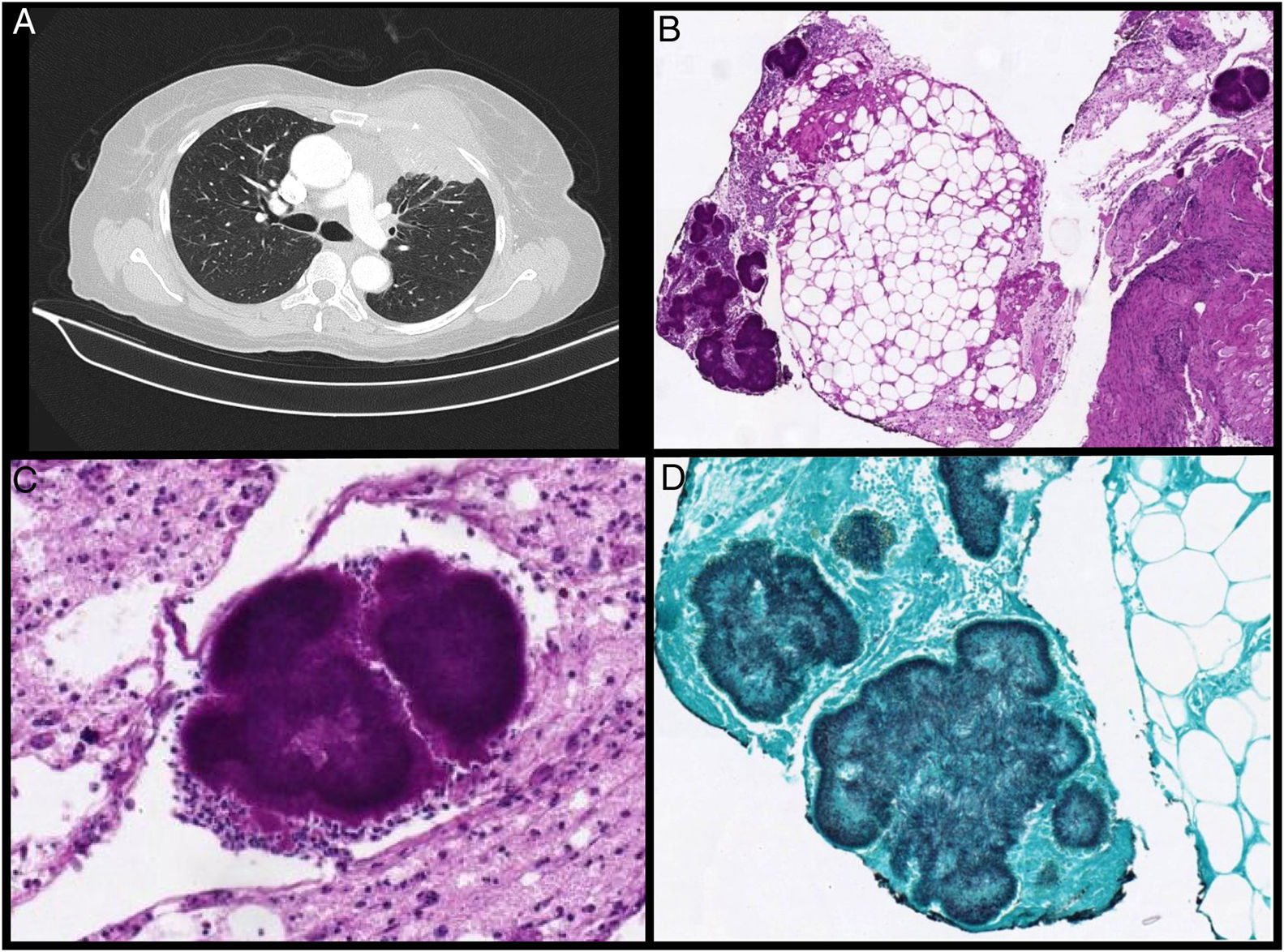
 Large mass in the left anterior chest wall affecting the minor and major pectoral muscles with underlying subpleural pulmonary parenchymal consolidation. The lung lesion contains an air bronchogram. (B) Histopathological sample (40×) stained with PAS showing a fragment of fibroadipose tissue with mixed inflammatory reaction and actinomycotic granules (Actinomyces colonies). (C) Image of an actinomycotic granule with PAS staining, surrounded by inflammatory infiltrate with abundant polymorphonuclear leukocytes (400×). (D) Anatomopathological sample processed with silver methenamine stain, revealing Actinomyces filaments in a palisade arrangement around the granule (400×).")
(A) Large mass in the left anterior chest wall affecting the minor and major pectoral muscles with underlying subpleural pulmonary parenchymal consolidation. The lung lesion contains an air bronchogram. (B) Histopathological sample (40×) stained with PAS showing a fragment of fibroadipose tissue with mixed inflammatory reaction and actinomycotic granules (Actinomyces colonies). (C) Image of an actinomycotic granule with PAS staining, surrounded by inflammatory infiltrate with abundant polymorphonuclear leukocytes (400×). (D) Anatomopathological sample processed with silver methenamine stain, revealing Actinomyces filaments in a palisade arrangement around the granule (400×).
Pulmonary actinomycosis is a chronic suppurative infection caused by gram-positive bacilli of the Actinomyces group that form part of our usual flora. Since clinical and radiological signs are non-specific, and may suggest a neoplastic process, diagnosis is usually delayed and generally obtained from a histopathological study. Treatment of choice is a 6–12 month course of penicillin or amoxicillin.
Actinomycosis should be taken into account when determining the etiology of atypical pneumonias in immunocompetent patients1,2.
Please cite this article as: Ramos AU, Hernández DM, Gallizo JA. Actinomicosis pulmonar en forma de masa pétrea pectoral. Arch Bronconeumol. 2020;56:592.


