


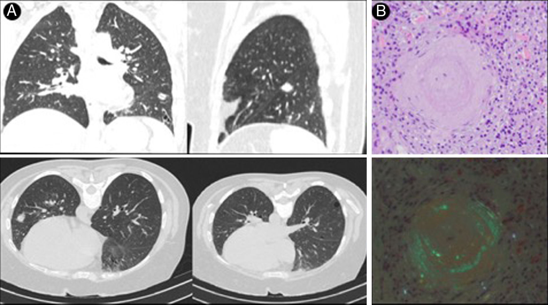
A 45-year-old woman with a history of mucosa-associated lymphoid tissue non-Hodgkin lymphoma, with complete response after treatment. A follow-up computed tomography (CT) scan showed multiple nodular lesions in a subpleural location and pulmonary cysts (Fig. 1A) with low uptake on positron emission tomography-computed tomography (PET-CT) (SUVmax. 1.3). Incidental findings on mammography included a breast node and a skin lesion in the lower limb that was biopsied, giving a diagnosis of AL amyloidosis (kappa-light chain). In view of suspected pulmonary nodular amyloidosis (PNA), a CT-guided core needle biopsy was performed that showed Congo red-positive material (Fig. 1B) and intensely positive kappa light chains in the immunohistochemical study, giving a diagnosis of systemic amyloidosis (AL) kappa-light chain.

PNA is an uncommon disease characterized by the presence of one or more amyloid tissue deposits in the lung. Primary amyloidosis is the most common, while systemic amyloidosis is rare. Patients are usually asymptomatic, and the disease is detected by an incidental finding in imaging studies showing well-defined nodules of subpleural distribution measuring 0.4−5 cm. Histological analysis revealing eosinophilic material stained with Congo red with apple green birefringence is necessary for diagnosis.1,2
Please cite this article as: Ruiz-Álvarez I, Gutiérrez Palacios AM, Rodríguez Díaz B. Amiloidosis nodular pulmonar, una causa infrecuente de nódulos pulmonares múltiples. Arch Bronconeumol. 2021;57:227.


