


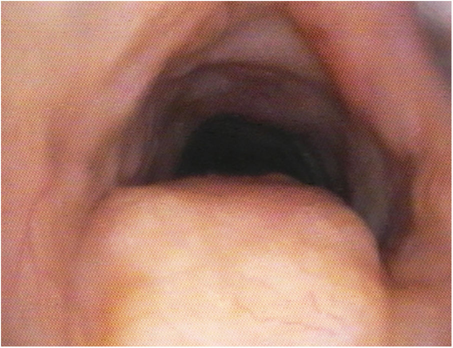
Our patient was an 80-year-old woman with no toxic habits or medical/surgical history of interest who underwent emergency appendectomy. Intubation was complicated by subglottic stenosis observed during direct laryngoscopy. The computed tomography (CT) scan showed nonspecific subglottic thickening of pseudonodular appearance (19 × 15 × 9 mm) in the posterior aspect of the proximal tracheal wall, partially compromising the airway. Flexible bronchoscopy (Appendix B, video and supplementary material) revealed a grayish-yellow lesion of granulomatous appearance with diffuse, concentric infiltration of the mucous membrane obstructing 30%–40% of the tracheal lumen (Fig. 1). Histology showed deposits of amorphous material that took on a reddish color after exposure to Congo red staining, in addition to apple-green birefringence in polarized light, consistent with amyloid. Invasive intervention was ruled out in the absence of symptoms, and the patient was followed up as an outpatient.

Amyloidosis of the upper airway is a rare, uncommon disease, with very nonspecific symptoms,1 that can sometimes be easily confused with other more common entities such as asthma, COPD, or lung cancer.2 A comprehensive assessment including high-resolution CT to determine the type of pulmonary involvement is essential, and flexible bronchoscopy with biopsy should be performed for histopathological confirmation of the diagnosis.
Please cite this article as: Felipe Montiel A, et al. Obstrucción no maligna de vía aérea superior: raro caso de amiloidosis traqueal. Arch Bronconeumol. 2020;56:252.


