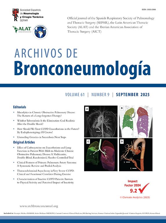

A 45-year-old healthy man was referred to Pulmonology outpatient department with persistent cough and wheezing, without other symptoms. Chest radiograph showed a homogeneous mass near the right cardiophrenic angle. Thoracic CT scan revealed a 15cm×8.2cm heterogeneous, vascular-like, fusiform lesion located under atelectatic, sequestrated lung parenchyma below the lower right lung lobe (Fig. 1A and B). The lesion had signs of mural thrombosis (Fig. 1C) and communicated proximally with an anomalous arterial branch from systemic circulation and distally with sequestrated lung parenchyma, with venous drainage into the pulmonary veins (Fig. 1C and D).
![(A) Thorax CT scan coronal images in two different planes (parenchymal window) showing atelectatic sequestrated lung (black arrows), pleural line totally separating normal lung parenchyma from underlying sequestrated lung (white arrowheads with black outline) and the lumen of the aneurysm (black arrowhead with grey outline). (B) Thorax CT scan axial image (parenchymal window) showing atelectatic sequestrated lung (black arrows) and the lumen of the aneurysm (black arrowhead with grey outline). (C) On the left – thorax CT scan coronal image (oblique maximum intensity projection [MIP] image) showing part of the aberrant systemic artery supplying sequestrated lung: an anomalous small arterial branch stemming from the posterior aspect of the descending thoracic aorta (black arrowhead with grey outline on the right), the lumen of the vessel inside the aneurysm (black arrowhead with grey outline on the left) and a peripheral hypodense component inside the aneurysm, suggesting mural thrombosis (black asterisk). On the right – volume rendered CT scan of the lumen of the same aberrant artery. Note that black arrowheads with grey outline roughly point to the same parts of the artery which are marked on the left part of figure C. White arrows mark the distal end of the vessel towards sequestrated lung parenchyma. (D) Thorax CT scan coronal images (oblique MIP image) showing other details regarding vascularization of sequestrated lung. Note that white arrows roughly point to the same parts of the artery which are marked on the right part of figure C. Grey arrowheads mark two almost parallel veins stemming from sequestrated lung parenchyma, converging towards pulmonary veins and finally draining to the left atrium.](https://static.elsevier.es/multimedia/03002896/0000005500000006/v1_201906140919/S0300289618304071/v1_201906140919/en/main.assets/gr1.jpeg?xkr=ue/ImdikoIMrsJoerZ+w9zfHQrK7O1GixshFj/RYvvhfIeFLIshOrwnoGDwJytw3sGm8bijV+TbKpnl+SctEd7MLy9rcXGgQK4WYHMZRma6r6i0mfu8CTNxKj4CMCxW+rrqjeqn3rVpyRZUdBL9FtVMZWA3X7/VnTayeO3OF1WHNoEFpeGyDqbCCdiFJ6uWQ49sgN9q0kz2zm/PyrOkUpz1FZl1cfSXfD5g2n4LT9FKXRqB5uBRcrFp0slbkvNci69ch7rndJgfZsITqB7Hui+GLSegd3zVTuKMGYR10aRY= "(A) Thorax CT scan coronal images in two different planes (parenchymal window) showing atelectatic sequestrated lung (black arrows), pleural line totally separating normal lung parenchyma from underlying sequestrated lung (white arrowheads with black outline) and the lumen of the aneurysm (black arrowhead with grey outline). (B) Thorax CT scan axial image (parenchymal window) showing atelectatic sequestrated lung (black arrows) and the lumen of the aneurysm (black arrowhead with grey outline). (C) On the left – thorax CT scan coronal image (oblique maximum intensity projection [MIP] image) showing part of the aberrant systemic artery supplying sequestrated lung: an anomalous small arterial branch stemming from the posterior aspect of the descending thoracic aorta (black arrowhead with grey outline on the right), the lumen of the vessel inside the aneurysm (black arrowhead with grey outline on the left) and a peripheral hypodense component inside the aneurysm, suggesting mural thrombosis (black asterisk). On the right – volume rendered CT scan of the lumen of the same aberrant artery. Note that black arrowheads with grey outline roughly point to the same parts of the artery which are marked on the left part of figure C. White arrows mark the distal end of the vessel towards sequestrated lung parenchyma. (D) Thorax CT scan coronal images (oblique MIP image) showing other details regarding vascularization of sequestrated lung. Note that white arrows roughly point to the same parts of the artery which are marked on the right part of figure C. Grey arrowheads mark two almost parallel veins stemming from sequestrated lung parenchyma, converging towards pulmonary veins and finally draining to the left atrium.")
(A) Thorax CT scan coronal images in two different planes (parenchymal window) showing atelectatic sequestrated lung (black arrows), pleural line totally separating normal lung parenchyma from underlying sequestrated lung (white arrowheads with black outline) and the lumen of the aneurysm (black arrowhead with grey outline). (B) Thorax CT scan axial image (parenchymal window) showing atelectatic sequestrated lung (black arrows) and the lumen of the aneurysm (black arrowhead with grey outline). (C) On the left – thorax CT scan coronal image (oblique maximum intensity projection [MIP] image) showing part of the aberrant systemic artery supplying sequestrated lung: an anomalous small arterial branch stemming from the posterior aspect of the descending thoracic aorta (black arrowhead with grey outline on the right), the lumen of the vessel inside the aneurysm (black arrowhead with grey outline on the left) and a peripheral hypodense component inside the aneurysm, suggesting mural thrombosis (black asterisk). On the right – volume rendered CT scan of the lumen of the same aberrant artery. Note that black arrowheads with grey outline roughly point to the same parts of the artery which are marked on the left part of figure C. White arrows mark the distal end of the vessel towards sequestrated lung parenchyma. (D) Thorax CT scan coronal images (oblique MIP image) showing other details regarding vascularization of sequestrated lung. Note that white arrows roughly point to the same parts of the artery which are marked on the right part of figure C. Grey arrowheads mark two almost parallel veins stemming from sequestrated lung parenchyma, converging towards pulmonary veins and finally draining to the left atrium.
Bilateral sequential thoracotomy with proximal ligation of the anomalous systemic artery, surgical repair of the aneurysm and wedge resection of adjacent sequestrated lung was performed with no postoperative complications. Histopathology supported the diagnosis of extralobar intrathoracic pulmonary sequestration supplied by an anomalous aneurysmatic artery.
Post-operatory thoracic CT scan was unremarkable. The patient remains asymptomatic.
Pulmonary sequestration consists of aberrant, congenital formation of segmental lung tissue unconnected with the tracheobronchial tree and pulmonary arteries, supplied by a systemic artery. Extralobar sequestration is the least common type and has male predilection.1,2 In this case, the risk of spontaneous aneurysm rupture and the high surgical risk were major concerns.
We would like to thank José Miranda, MD for the decisive role in the surgical treatment of this patient. We also thank Manuela França, MD, PhD for the thorough revision of CT scan images of this patient.


