Journal Information
Share
Share
Download PDF
More article options
Visits
55
Original Article
Full text access
Uncorrected Proof. Available online 17 July 2024
Efficacy and Safety of Platelet-Rich Plasma on Bronchopleural Fistula: A Pilot Prospective Cohort Study
Visits
55
Yongshun Yea,b,1, Tingting Xua,1, Jinxia Lina,1, Yongna Caia, Zhuquan Sua, Liya Luc, Yu Chena, Changgao Zhonga, Chunli Tanga, Weiquan Xiaoa, Haojie Liaob, Shiyue Lia,2,
Corresponding author
lishiyue@188.com

Corresponding authors.
, Xiaobo Chena,2,
Corresponding author
xiaobo-win@163.com

Corresponding authors.
a Guangzhou Institute of Respiratory Health, State Key Laboratory of Respiratory Disease, National Clinical Research Center for Respiratory Disease, National Center for Respiratory Medicine, The First Affiliated Hospital of Guangzhou Medical University, Guangzhou 510120, China
b Huizhou Central People's Hospital, Huizhou 516000, China
c Department of Anesthesiology Department, The First Affiliated Hospital of Guangzhou Medical University, 151 Yan Jiang Rd, Guangzhou 510120, China
This item has received
Article information
Abstract
Full Text
Bibliography
Download PDF
Statistics
Figures (4)
Show moreShow less
Tables (3)
Table 1. Patient demographics and baseline characteristics of bronchopleural fistulas.
Table 2. Details of PRP treatment and the therapeutic effect of PRP.
Table 3. Summary of device-related adverse events during the treatment period and follow-up period.
Show moreShow less
Abstract
Introduction

Bronchopleural fistula (BPF) is associated with high morbidity and mortality rates in patients undergoing pulmonary resections. Surgery, bronchoscopy, and conservative management have their limitations for small fistulas. Platelet-rich plasma (PRP) has regenerative properties, which might be efficient in enhancing tissue recovery and repairing small BPF. This study aimed to investigate efficacy and safety of PRP on BPF.

Methods

This is a pilot prospective cohort study. Patients whose fistulas smaller than 4mm were enrolled in this study, treated with PRP under bronchoscopy and followed up at 2 weeks and 4–6 weeks after the last PRP treatment. The cure rate, improvement rate and ineffectiveness rate were investigated. The severity of respiratory symptoms was evaluated by modified Medical Research Council dyspnea scale (mMRC) and COPD Assessment Test (CAT). The recurrence of fistula, new infection and mortality rate were examined. Adverse events were documented to explore the safety profile of PRP therapy.
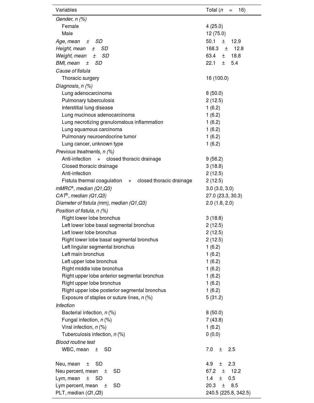
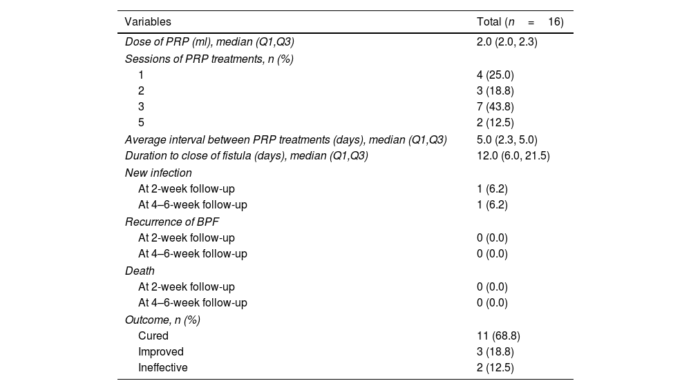
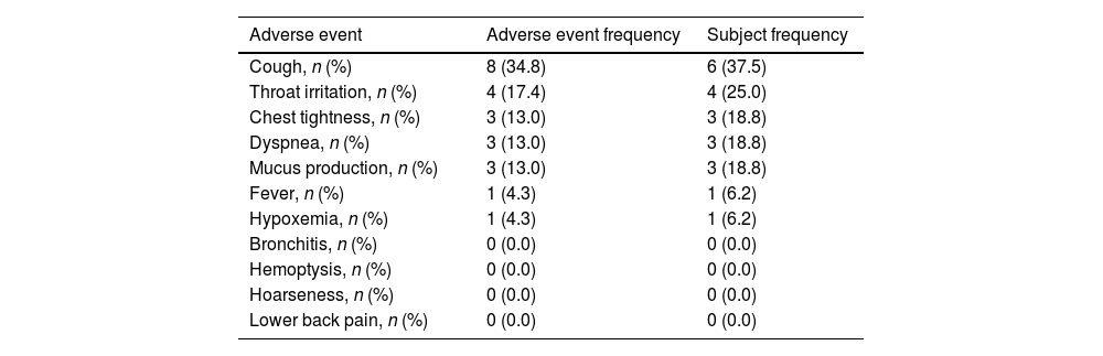
Results

A total of 16 patients (mean age, 50.1 years) met the eligibility criteria. The median time from the first PRP treatment to the closure of the fistula was 12.0 (IQR 6.0, 21.5) days. Our findings indicate an effectiveness rate of 87.6%, with 68.8% of cure and 18.8% of improvement, along with significant improvement of respiratory symptoms evaluated by mMRC (P<0.001) and CAT (P<0.001). No recurrent of fistulas, newly developed infection, or death was observed. Adverse events of the procedure were most mild (82.6%) and temporary.

Conclusions

PRP is a potential treatment for small BPF and is well tolerated.

Keywords:
Platelet-rich plasma
Bronchial fistula
Treatment outcome
Safety
Regenerative medicine
Full Text
Introduction

Bronchopleural fistula (BPF), an abnormal connection between the bronchial tree and pleural space, is a severe condition with high morbidity and mortality rates in patients, often seen after pulmonary resections due to lung cancer, tuberculosis, trauma, or radiation.1–3 The treatment strategy for BPF depends on the fistula size, the patient's condition, and the timing post-surgery.2 Surgical intervention is typically reserved for large fistulas and comes with high morbidity and mortality rates.1–4 In contrast, bronchoscopic techniques such as blockers, tissue adhesives, and stents are used for smaller fistulas but can still lead to recurrence and death.5–7 The complexity of treating BPF, especially small fistulas, highlights the need for further development of regenerative repair strategies.

Recent studies have highlighted the widespread application of platelet-rich plasma (PRP) in regenerative medicine. The enriched growth factors substantially promote tissue repair and regeneration across various fields such as orthopedics, dermatology, and wound healing. These studies underscore PRP's ability to expedite tissue recovery and mitigate inflammation.8,9 The use of autologous PRP significantly reduces the likelihood of immune rejection and adverse reactions, offering a notable advantage over traditional therapies. In our prior research, we successfully leveraged PRP therapy to address benign airway stenosis, effectively mitigating the risk of subsequent narrowing.10 PRP has unveiled its regenerative and reparative capabilities for airway conditions. The regenerative capabilities of PRP have sparked an increased interest in managing complex conditions, including BPF.

This pilot prospective cohort study aims to evaluate the efficacy and safety of PRP for small BPF, and endeavors to provide a novel therapeutic avenue for BPF patients, potentially improving clinical outcomes and quality of life. By rigorously analyzing PRP's utility in a clinical setting, this research intends to provide valuable insights into its potential as a novel treatment for BPF, thereby advancing regenerative medicine applications and informing future therapeutic strategies.

Material and methodStudy design

This nonrandomized, prospective study enrolled 16 BPF patients at the First Affiliated Hospital of Guangzhou Medical University from April 16, 2022, to December 20, 2023, with follow-up until January 25, 2024. The protocol was approved by the Ethics Committee of the First Affiliated Hospital of Guangzhou Medical University (No. 2021-118). Written, informed consent was obtained from all subjects. The trial was registered at Clinicaltrials.gov (Clinicaltrials.gov number, NCT05304897).

Study participants

The selection and follow-up processes are depicted in the flowchart (Fig. 1). The eligibility criteria included subjects aged between 18 and 75, diagnosed with BPF with sizes less than 4mm, and willing to accept PRP treatment. Subjects with fistulas larger than 4mm, those who cannot tolerate bronchoscopy due to severe cardiac disease or other major comorbidities, and pregnant or breastfeeding women were excluded.

Fig. 1.

The flowchart of this study.

(0.12MB).
Preoperative evaluation

At enrollment, demographics, the cause of BPF, diagnosis, and prior treatments were recorded. Dyspnea severity was measured using the modified Medical Research Council dyspnea scale (mMRC), and respiratory symptoms and quality of life were assessed with the COPD Assessment Test (CAT). Fistula position, diameter, and any exposed sutures or staples were evaluated via bronchoscopy and chest CT. Infection presence and type were identified through etiological examination of pleural drainage fluid.

Anesthesia method

All patients received sedation and analgesia in the operating room with spontaneous respiration preserved. They were pre-medicated with intravenous dexmedetomidine (0.2μg/kg) 15min before anesthesia. Anesthesia induction was intravenous sufentanil (0.2μg/kg) and propofol (13μg/ml). Anesthesia during surgery was maintained with propofol and remifentanil (0.05–0.08μg/kg/min), with a Bispectral Index (BIS) of 60–80 maintained and continuous vital signs monitoring.

PRP extraction

Initially, about 15ml of blood was collected from the patient's antecubital vein into a sodium citrate tube. The PRP was isolated using the Arthrex ACP system (ABS-10014, Arthrex, Inc., USA), a PRP separation device, according to manufacturer's instructions.

Surgical procedure

Prior to PRP treatment, necrotic tissue, exposed sutures, and anastomotic staples near the fistula were removed with a flexible bronchoscope (BF-260, Olympus, Japan), and the area was cleansed with saline and suctioned. PRP was administered around the fistula via the bronchoscope, with each point receiving about 0.2ml of PRP, placed at least 1mm from the fistula's edge. Total PRP per session was 2–3ml, depending on the fistula's size and condition. A coagulant mix (thrombin 2000ui+3% calcium chloride 10ml) at 0.3ml was then applied to the area to promote PRP clotting and cover the fistula.

Standard for closure of fistula and criteria for continuing or terminating PRP treatment

Multiple PRP treatments have been shown to be superior to single PRP treatments.11,12 To better evaluate the efficacy, we have established the following criteria of closure: (1) no air leak for 7 days after any PRP; (2) lung re-expansion on CT/X-ray at least 7 days post-PRP; or (3) closed fistula on bronchoscopy at least 7 days post-PRP. PRP treatment protocols were: (1) terminate PRP if closure criteria met, with possible additional treatment for consolidation upon physician's decision; (2) continue PRP if criteria not met, with a total of no more than 5 sessions per individual case; (3) stop PRP if patient opts out. All patients will undergo efficacy evaluation after completing treatment, and the definition of the evaluation results can be found in the “Outcomes” section.

Post-treatment monitoring and evaluation

Subjects had follow-ups at 2 weeks and 4–6 weeks post-last PRP treatment for re-evaluation. BPF recurrence and infections were initially assessed via symptoms, with chest CT and microbiological tests on drainage fluid conducted as needed. mMRC and CAT scores were re-assessed at these follow-ups. Safety was monitored by recording adverse events post-treatment and through weekly telephone check-ups for 6 weeks. Adverse events were classified by investigators as mild (no activity disruption), moderate (interfered with daily activities, manageable with symptomatic treatment), or severe (incapacitating, requiring medical intervention).

Outcomes

The primary outcome was the cure rate of BPF. Cure was defined as achieving the criteria for fistula closure and no recurrence during a follow-up of 4 weeks or more. Improvement was defined as a reduction of more than 50% in the diameter of the fistula under bronchoscopy or a decrease in the gas leakage from the drainage tube at the last follow-up. Ineffectiveness was defined as no reduction or less than 50% reduction in the diameter of the fistula under bronchoscopy and with no significant change in the gas leakage from the drainage tube at the last follow-up. The secondary outcomes were the alleviation or aggravation of dyspnea assessed by the mMRC scale, and change of respiratory symptoms and quality of life evaluated by the CAT.

Statistics

This study was designed to evaluate the safety and efficacy of PRP in patients with BPF. Statistical analyses of efficacy measures were performed for information. Categorical variables were presented as frequency (percentage), while continuous variables were expressed as mean±SD or median (interquartile range). The changes in mMRC and CAT were analyzed using repeated measures ANOVA. The Statistical analysis was performed by using R software (v 4.1.2).

ResultsPatient demographics

A total of 33 patients were enrolled for evaluation, among which 17 subjects were excluded (13 with diameter of fistula larger than 4mm, 3 withdrew consent, 1 with severe cardiac diseases). In total, 16 patients were enrolled, with 12 (75.0%) males, a mean age of 50.1 (SD 12.9) years, and a mean BMI of 22.1 (SD 5.4)kg/m2. Baseline subject characteristics are listed in Table 1. All patients developed BPF due to surgical procedures. Diagnoses included lung adenocarcinoma (50.0%), pulmonary tuberculosis (12.5%), and other lung conditions. Previous treatment consisted of the combination of anti-infection treatment and closed thoracic drainage (56.2%), closed thoracic drainage (18.8%), anti-infection treatment (12.5%), the combination of fistula thermal coagulation and closed thoracic drainage (12.5%). Bronchoscopic procedures revealed that the median diameter of the fistulas was 2.0 (IQR 1.8, 2.0)mm. The location of the BPF varied among patients. In 5 (31.2%) patients, exposed staples or sutures were observed under the bronchoscope. Microbiological examination of the closed thoracic drainage fluid revealed that 8 (50.0%) patients had bacterial infections, 7 (43.8%) had fungal infections, 1 (6.2%) had a viral infection, and no patients had tuberculosis infections. The median platelet count was 240.5 (IQR 225.8, 342.5).

Table 1.

Patient demographics and baseline characteristics of bronchopleural fistulas.

Variables  Total (n=16) 
Gender, n (%)
Female  4 (25.0) 
Male  12 (75.0) 
Age, mean±SD  50.1±12.9 
Height, mean±SD  168.3±12.8 
Weight, mean±SD  63.4±18.8 
BMI, mean±SD  22.1±5.4 
Cause of fistula
Thoracic surgery  16 (100.0) 
Diagnosis, n (%)
Lung adenocarcinoma  8 (50.0) 
Pulmonary tuberculosis  2 (12.5) 
Interstitial lung disease  1 (6.2) 
Lung mucinous adenocarcinoma  1 (6.2) 
Lung necrotizing granulomatous inflammation  1 (6.2) 
Lung squamous carcinoma  1 (6.2) 
Pulmonary neuroendocrine tumor  1 (6.2) 
Lung cancer, unknown type  1 (6.2) 
Previous treatments, n (%)
Anti-infection+closed thoracic drainage  9 (56.2) 
Closed thoracic drainage  3 (18.8) 
Anti-infection  2 (12.5) 
Fistula thermal coagulation+closed thoracic drainage  2 (12.5) 
mMRCa, median (Q1,Q3)  3.0 (3.0, 3.0) 
CATb, median (Q1,Q3)  27.0 (23.3, 30.3) 
Diameter of fistula (mm), median (Q1,Q3)  2.0 (1.8, 2.0) 
Position of fistula, n (%)
Right lower lobe bronchus  3 (18.8) 
Left lower lobe basal segmental bronchus  2 (12.5) 
Left lower lobe bronchus  2 (12.5) 
Right lower lobe basal segmental bronchus  2 (12.5) 
Left lingular segmental bronchus  1 (6.2) 
Left main bronchus  1 (6.2) 
Left upper lobe bronchus  1 (6.2) 
Right middle lobe bronchus  1 (6.2) 
Right upper lobe anterior segmental bronchus  1 (6.2) 
Right upper lobe bronchus  1 (6.2) 
Right upper lobe posterior segmental bronchus  1 (6.2) 
Exposure of staples or suture lines, n (%)  5 (31.2) 
Infection
Bacterial infection, n (%)  8 (50.0) 
Fungal infection, n (%)  7 (43.8) 
Viral infection, n (%)  1 (6.2) 
Tuberculosis infection, n (%)  0 (0.0) 
Blood routine test
WBC, mean±SD  7.0±2.5 
Variables  Total (n=16) 
Neu, mean±SD  4.9±2.3 
Neu percent, mean±SD  67.2±12.2 
Lym, mean±SD  1.4±0.5 
Lym percent, mean±SD  20.3±8.5 
PLT, median (Q1,Q3)  240.5 (225.8, 342.5) 
a

Modified Medical Research Council dyspnea scale.

b

COPD Assessment Test.

PRP treatment

All bronchoscopic procedures were completed in less than 30min. After injecting PRP around the fistula site, immediate closure of the fistula due to swelling of the surrounding tissue was observed under bronchoscopy, accompanied by the cessation of air discharge from the closed thoracic drainage tube. After the acute phase of tissue swelling subsided, air discharge from the closed thoracic drainage tube could reoccur, accompanied by the recurrence of fistula observed in subsequent bronchoscopic examinations. In these cases, further PRP treatment was necessary. In cases who were cured, healing scars at the origin fistula could be observed under bronchoscopy. Chest CT, chest X-ray and alterations of macroscopic appearance under bronchoscopy before and after PRP treatment of a typical case are available in the supplementary materials. In total, 41 bronchoscopic procedures were performed on 16 patients, with one session in 4 (25%) patients, 2 sessions in 3 (18.8%) patients, 3 sessions in 7 (43.8%) patients, and 5 sessions in 2 (12.5%) patients. The PRP dosage used in each treatment was 2.0 (IQR 2.0, 2.3) ml. The median interval between each PRP treatment was 5 (IQR 2.3, 5.0) days (Table 2).

Table 2.

Details of PRP treatment and the therapeutic effect of PRP.

Variables  Total (n=16) 
Dose of PRP (ml), median (Q1,Q3)  2.0 (2.0, 2.3) 
Sessions of PRP treatments, n (%)
4 (25.0) 
3 (18.8) 
7 (43.8) 
2 (12.5) 
Average interval between PRP treatments (days), median (Q1,Q3)  5.0 (2.3, 5.0) 
Duration to close of fistula (days), median (Q1,Q3)  12.0 (6.0, 21.5) 
New infection
At 2-week follow-up  1 (6.2) 
At 4–6-week follow-up  1 (6.2) 
Recurrence of BPF
At 2-week follow-up  0 (0.0) 
At 4–6-week follow-up  0 (0.0) 
Death
At 2-week follow-up  0 (0.0) 
At 4–6-week follow-up  0 (0.0) 
Outcome, n (%)
Cured  11 (68.8) 
Improved  3 (18.8) 
Ineffective  2 (12.5) 
Efficacy outcomesCure rate

The median time from the first PRP treatment to the closure of the fistula was 12.0 (IQR 6.0, 21.5) days. After PRP treatment, 11 (68.8%) patients were cured, 3 (18.8%) showed improvement, and 2 (12.5%) gained no efficacy from the treatment (Table 2).

Severity of symptoms

It was found that before PRP treatment, the median mMRC score was 3.0 (IQR 3.0, 3.0). Two weeks (P<0.001) and 4–6 weeks (P<0.001) after the last PRP treatment, the mMRC scores significantly decreased, with median mMRC scores of 1.0 (IQR 0.0, 1.0) and 0.0 (IQR 0.0, 0.3), respectively (Fig. 2). CAT revealed that before PRP therapy, the median CAT score was 27.0 (IQR 23.3, 30.3). There was a significant reduction in CAT scores two weeks (P<0.001) and 4–6 weeks (P<0.001) following the final PRP session. Specifically, median CAT scores dropped to 3.0 (IQR 2.0, 4.3) 2 weeks after and further to 1.5 (IQR 0.0, 3.0) 4–6 weeks after the last treatment, indicating substantial improvements in patients’ respiratory symptoms and quality of life (Fig. 3).

Fig. 2.

The mMRC at baseline, 2 weeks after the last PRP treatment, and 4–6 weeks after the last PRP treatment.

(0.12MB).
Fig. 3.

The CAT at baseline, 2 weeks after the last PRP treatment, and 4–6 weeks after the last PRP treatment.

(0.19MB).
Recurrence of BPF, new infection, and death

At the 2-week and 4–6-week follow-up visits, no recurrence of fistula was observed. One (6.2%) patient was found to have a newly developed tuberculosis infection in the thoracic cavity drainage fluid during the follow-up visits at 2 weeks and 4–6 weeks. However, this patient had obsolete lesions of tuberculosis in the lungs before PRP treatment, and the infection was considered unrelated to the PRP treatment or procedure. Throughout the study, there were no deaths among the participants.

Safety of PRP

For the safety analysis, all symptoms were documented, resulting in a total of 29 adverse events throughout the monitoring timeframe. Out of these, 23 were attributed to either the device or the procedure itself, essentially being identified as side effects. Table 3 details the characteristics of these side effects that exclusively occurred within the treatment and follow-up phases. The most common side effects involved airway irritation, which manifested as increased coughing and throat irritation. The onset of these symptoms typically occurred within 1.0 (IQR 0.0, 2.0) days, and resolution was usually observed in 2.0 (IQR 1.0, 2.0) days following the latest bronchoscopy. Among these device- and/or procedure-related side effects, 15 (65.2%) resolved spontaneously and 8 (34.8%) resolved after symptomatic treatment. Neither the frequency nor the intensity of side effects escalated with additional treatment sessions. In this study, most side effects were classified as mild, accounting for 19 (82.6%) of the incidents, while the rest were moderate, totaling 4 (17.4%). There were no reports of severe side effects.

Table 3.

Summary of device-related adverse events during the treatment period and follow-up period.

Adverse event  Adverse event frequency  Subject frequency 
Cough, n (%)  8 (34.8)  6 (37.5) 
Throat irritation, n (%)  4 (17.4)  4 (25.0) 
Chest tightness, n (%)  3 (13.0)  3 (18.8) 
Dyspnea, n (%)  3 (13.0)  3 (18.8) 
Mucus production, n (%)  3 (13.0)  3 (18.8) 
Fever, n (%)  1 (4.3)  1 (6.2) 
Hypoxemia, n (%)  1 (4.3)  1 (6.2) 
Bronchitis, n (%)  0 (0.0)  0 (0.0) 
Hemoptysis, n (%)  0 (0.0)  0 (0.0) 
Hoarseness, n (%)  0 (0.0)  0 (0.0) 
Lower back pain, n (%)  0 (0.0)  0 (0.0) 

A total of 23 events were reported.

Sixteen subjects experienced one or more reported events.

Discussion

This pilot prospective cohort study revealed a promising potential for PRP therapy in small BPF, a condition known for its challenging management, high morbidity, and mortality, providing a theoretical basis for its subsequent clinical application. Our findings indicate an effectiveness rate of 87.6%, with 68.8% cure and 18.8% improvement and an absence of recurrence of BPF and new infection during follow-up, accompanied by significant improvement of respiratory symptoms. Furthermore, a favorable safety profile was observed in this study.

BPF, commonly following lobectomy, lacks standardized treatment. Main methods include surgical and bronchoscopic interventions. Surgical repair, primarily using pedicled intercostal muscle flaps, has a high success rate of 95.3%.13 However, Hollaus et al. reported severe complications in 7 patients (an average fistula size of 3.43mm) treated by pedicled intercostal muscle flaps, including skin flap necrosis in 1 patient, and death in 1 case caused by aspiration pneumonia 38 days post-thoracotomy.14 Alternatively, endoscopic treatment offers minimal invasiveness and flexibility, combining multiple techniques with success rates up to 80%.5 Devices like the Amplatzer occluders and Amplatzer vascular plug effectively seal BPF, achieving immediate success in 96% of cases.15 However, the application is limited by the size of the fistulas, with smaller ones being challenging to repair with occluders.16

PRP therapy under endoscopic guidance is an emerging treatment for BPF. PRP is rich in growth factors and cytokines like platelet-derived growth factor (PDGF), transforming growth factor-β (TGF-β), vascular endothelial growth factor (VEGF), and fibroblast growth factor (FGF), which are crucial for tissue repair. Its regenerative properties have been effectively applied in healing bone,17,18 cartilage,19,20 and skin injuries,21 among others. PRP has shown promise in airway regeneration, with cases reporting complete healing in bronchial anastomotic fistulas post-lung transplantation.22 Our team refined PRP formulations and focused on the active component ARF, demonstrating its effectiveness in preventing restenosis in refractory benign airway stenosis.10 Another study conducted by Wu et al. explored PRP for BPF in 3 cases. All 3 cases were cured, though the treatment duration (up to 124 days) and additional interventions clouded PRP's sole effects.23 In our structured study using endoscopic PRP application, we established a rigorous protocol, achieving an overall efficacy of 87.6%, with a cure rate of 68.8% and improvement rate of 18.8%. This approach underscores PRP's therapeutic potential in airway repair. In our study, we observed immediate swelling of the surrounding tissue after PRP infection. However, PRP offers additional benefits beyond just causing tissue swelling. The growth factors and cytokines in it can reduce inflammation and promote tissue regeneration and healing, leading to permanent close of fistula.

PRP is considered safe for airway applications. A systematic review of 21 studies on PRP treatment for osteoarthritis identified only mild adverse reactions such as pain, redness, and swelling.24 In airway applications, Wu et al. found no adverse reactions in three cases of tracheobronchial fistula treated with PRP.23 Our research also indicates that potential adverse reactions in airways, including cough, dyspnea, and chest discomfort, are typically short-lived, further confirming PRP's safety in treating conditions like BPF.10

PRP's effectiveness can vary in treating orthopedic diseases, dental issues, and chronic dermatoses,25,26 often due to factors like PRP quality, application methods, patient characteristics, and treatment timing.27 In this study, we did not observe that the type of surgery, location, or disease affected the efficacy of PRP treatment. Among the patients in our study, both cases of treatment failure were observed in patients who had undergone surgery for left lung cancer. The age, fistula size, and fistula location of these two patients were similar to those of BPF patients who were cured with PRP therapy. Upon reviewing the surgical videos, we noted that local tissue scarring around the fistula might be one of the factors affecting the action of PRP growth factors. Evidence suggests that the structural characteristics and microenvironment of the wound influence the success of regenerative therapies.26 However, this hypothesis requires further validation through larger clinical studies.

The presence of multiple BPF is another circumstance that cannot be ignored. In our study of multiple BPFs, two patients had dual fistulas, including one at the distal bronchi inaccessible by bronchoscopy. We used bronchoscopy to check the proximal fistulas’ closure, but final chest CTs showed residual cavities. Given the stability of the distal fistula and potential risks to lung function, no further interventions were pursued. This highlights the challenges in treating multiple BPFs and the need for personalized, multi-modal treatment approaches when fistulas are unreachable by standard endoscopic methods.

Recurrence of BPF after treatment is also a common concern.4,29 In our current study, we observed no recurrence of fistula, indicating initial success. However, the potential for recurrence may increase with extended observation periods and larger sample sizes. The efficacy of further PRP in patients with recurrence of fistula remains an area for future exploration to fully understand and optimize BPF management strategies.

This study has several limitations. The first one is the absence of a control group and a small sample size. Despite this, significant improvements in cure rates and respiratory symptoms were observed. These results are promising and suggest that PRP treatment might offer benefits beyond those achievable with occluders, bronchial stents, and conservative approaches, warranting further research. Second, 4 patients, of whom 2 had outcomes of ineffectiveness and 2 showed improvement, declined further PRP treatments due to concerns about the risks associated with repeated anesthesia and bronchoscopy. This refusal may have led to an underestimation of PRP's therapeutic efficacy in the study. Finally, this study does not address potential long-term safety issues like recurrence of BPF, new infections, or death. These issues are intended to be addressed through more rigorous study designs, which are multicenter randomized controlled trials and extended follow-up periods.

Conclusion

PRP therapy had significant potential in managing small BPF, with an effectiveness rate of around 4/5, along with notable improvements in respiratory symptoms. The absence of BPF recurrence or new infections during follow-up and few adverse events further underscored efficacy and safety of PRP. These findings laid a solid foundation for future clinical applications of PRP in small BPF treatment, suggesting a promising avenue for addressing this complex condition.

Ethics approval and consent to participate

The protocol was approved by the Ethics Committee of the First Affiliated Hospital of Guangzhou Medical University (No. 2021-118). The trial was registered at Clinicaltrials.gov (Clinicaltrials.gov number, NCT05304897).

Funding

This study was supported by the grant of State Key Laboratory of Respiratory Disease (SKLRD-Z-202201), Guangdong Province Key Field Research and Development Program (2023B1111050006), Innovation team of respiratory diseases and regenerative medicine (2021KCXTD028) and Clinical High-tech Project of Guangzhou (2023C-GX04). The funders had no role in study design, data collection and analysis, decision to publish, or preparation of the manuscript.

Authors’ contributions

All authors participated in manuscript writing and editing. Conception and design: Xiaobo Chen, Shiyue Li, Tingting Xu, Yongshun Ye. Recruiting subjects and acquisition of data: Xiaobo Chen, Jinxia Lin, Tingting Xu, Yongshun Ye, Yongna Cai, Zhuquan Su, Liya Lu, Yu Chen, Changgao Zhong, Chunli Tang, Weiquan Xiao. Statistical analysis: Tingting Xu, Yongshun Ye, Xiaobo Chen. Critical review and editing: Xiaobo Chen, Shiyue Li, Tingting Xu, Yongshun Ye. All authors read and approved the final manuscript.

Conflict of interest

The authors have no conflicts of interest to declare.

Availability of data and materials

The datasets used and/or analyzed during the current study are available from the corresponding author on reasonable request.

Uncited reference

28.

Acknowledgment

None.

References
[1]
M.H. Baumann, S.A. Sahn.
Medical management and therapy of bronchopleural fistulas in the mechanically ventilated patient.
Chest, 97 (1990), pp. 721-728
[2]
M. Lois, M. Noppen.
Bronchopleural fistulas: an overview of the problem with special focus on endoscopic management.
Chest, 128 (2005), pp. 3955-3965
[3]
H. Asamura, T. Naruke, R. Tsuchiya, T. Goya, H. Kondo, K. Suemasu.
Bronchopleural fistulas associated with lung cancer operations. Univariate and multivariate analysis of risk factors, management, and outcome.
J Thorac Cardiovasc Surg, 104 (1992), pp. 1456-1464
[4]
J.D. Puskas, D.J. Mathisen, H.C. Grillo, J.C. Wain, C.D. Wright, A.C. Moncure.
Treatment strategies for bronchopleural fistula.
J Thorac Cardiovasc Surg, 109 (1995), pp. 989-995
[5]
G. Cardillo, L. Carbone, F. Carleo, G. Galluccio, M. Di Martino, R. Giunti, et al.
The rationale for treatment of postresectional bronchopleural fistula: analysis of 52 patients.
Ann Thorac Surg, 100 (2015), pp. 251-257
[6]
R. Mao, P.Q. Ying, D. Xie, C.Y. Dai, J.Y. Zha, T. Chen, et al.
Conservative management of empyema-complicated post-lobectomy bronchopleural fistulas: experience of consecutive 13 cases in 9 years.
J Thorac Dis, 8 (2016), pp. 1577-1586
[7]
L. Jin, Y. Li.
Bronchoscopic interventions for bronchopleural fistulas.
Ther Adv Respir Dis, 17 (2023),
[8]
P. Samadi, M. Sheykhhasan, H.M. Khoshinani.
The use of platelet-rich plasma in aesthetic and regenerative medicine: a comprehensive review.
Aesthetic Plast Surg, 43 (2019), pp. 803-814
[9]
A. Lubkowska, B. Dolegowska, G. Banfi.
Growth factor content in PRP and their applicability in medicine.
J Biol Regul Homeost Agents, 26 (2012), pp. 3s-22s
[10]
X. Chen, W. Wang, Y. Ye, Y. Yang, D. Chen, R. He, et al.
The wound healing of autologous regenerative factor on recurrent benign airway stenosis: a canine experimental and pilot study.
Respiration, (2024), pp. 1-13
[11]
D.K. Chouhan, M.S. Dhillon, S. Patel, T. Bansal, A. Bhatia, H. Kanwat.
Multiple platelet-rich plasma injections versus single platelet-rich plasma injection in early osteoarthritis of the knee: an experimental study in a guinea pig model of early knee osteoarthritis.
Am J Sports Med, 47 (2019), pp. 2300-2307
[12]
Z. Jin, J. Fang, J. Wang, Y. Wang, Q. Song, X. Tian, et al.
Correction to: platelet-rich plasma combined with low-dose ultrashort wave therapy accelerates peripheral nerve regeneration by Zhu Y.
Tissue Eng Part A, 26 (2020), pp. 178-192
[13]
G. Stamatis, L. Freitag, M. Wencker, D. Greschuchna.
Omentopexy and muscle transposition: two alternative methods in the treatment of pleural empyema and mediastinitis.
Thorac Cardiovasc Surg, 42 (1994), pp. 225-232
[14]
P.H. Hollaus, M. Huber, F. Lax, P.N. Wurnig, G. Böhm, N.S. Pridun.
Closure of bronchopleural fistula after pneumonectomy with a pedicled intercostal muscle flap.
Eur J Cardiothorac Surg, 16 (1999), pp. 181-186
[15]
O. Fruchter, B.A. El Raouf, N. Abdel-Rahman, M. Saute, E. Bruckheimer, M.R. Kramer.
Efficacy of bronchoscopic closure of a bronchopleural fistula with amplatzer devices: long-term follow-up.
Respiration, 87 (2014), pp. 227-233
[16]
O. Fruchter, E. Bruckheimer, Y. Raviv, D. Rosengarten, M. Saute, M.R. Kramer.
Endobronchial closure of bronchopleural fistulas with Amplatzer vascular plug.
Eur J Cardiothorac Surg, 41 (2012), pp. 46-49
[17]
K. Kazakos, D.N. Lyras, D. Verettas, K. Tilkeridis, M. Tryfonidis.
The use of autologous PRP gel as an aid in the management of acute trauma wounds.
[18]
G. Milano, E. Sanna Passino, L. Deriu, G. Careddu, L. Manunta, A. Manunta, et al.
The effect of platelet rich plasma combined with microfractures on the treatment of chondral defects: an experimental study in a sheep model.
Osteoarthr Cartil, 18 (2010), pp. 971-980
[19]
Y. Sun, Y. Feng, C.Q. Zhang, S.B. Chen, X.G. Cheng.
The regenerative effect of platelet-rich plasma on healing in large osteochondral defects.
Int Orthop, 34 (2010), pp. 589-597
[20]
X. Xie, C. Zhang, R.S. Tuan.
Biology of platelet-rich plasma and its clinical application in cartilage repair.
Arthritis Res Ther, 16 (2014), pp. 204
[21]
P. Xu, Y. Wu, L. Zhou, Z. Yang, X. Zhang, X. Hu, et al.
Platelet-rich plasma accelerates skin wound healing by promoting re-epithelialization.
Burns Trauma, 8 (2020), pp. tkaa028
[22]
A. Siddique, B.N. Sabbah, T. Arabi, I.M. Shakir, R. Abdulqawi, K. AlKattan, et al.
Treatment of bronchial anastomotic fistula using autologous platelet-rich plasma post lung transplantation.
J Cardiothorac Surg, 17 (2022), pp. 204
[23]
M. Wu, H. Lin, L. Shi, L. Huang, Y. Zhuang, Y. Zeng.
Bronchoscopic treatment of tracheobronchial fistula with autologous platelet-rich plasma.
Ann Thorac Surg, 111 (2021), pp. e129-e131
[24]
S. Chavda, S.A. Rabbani, T. Wadhwa.
Role and effectiveness of intra-articular injection of hyaluronic acid in the treatment of knee osteoarthritis: a systematic review.
Cureus, 14 (2022), pp. e24503
[25]
N. Jakse, S. Tangl, R. Gilli, A. Berghold, M. Lorenzoni, A. Eskici, et al.
Influence of PRP on autogenous sinus grafts. An experimental study on sheep.
Clin Oral Implants Res, 14 (2003), pp. 578-583
[26]
J. Emer.
Platelet-rich plasma (PRP): current applications in dermatology.
Skin Therapy Lett, 24 (2019), pp. 1-6
[27]
P. Oneto, J. Etulain.
PRP in wound healing applications.
Platelets, 32 (2021), pp. 189-199
[28]
E. Fitzpatrick, O.J. Holland, J.J. Vanderlelie.
Ozone therapy for the treatment of chronic wounds: a systematic review.
Int Wound J, 15 (2018), pp. 633-644
[29]
G. Kalweit, P. Feindt, H. Huwer, I. Volkmer, E. Gams.
The pectoral muscle flaps in the treatment of bronchial stump fistula following pneumonectomy.
Eur J Cardiothorac Surg, 8 (1994), pp. 358-362

Contributed equally as co-first authors.

Contributed equally as co-corresponding authors.

Copyright © 2024. SEPAR
Archivos de Bronconeumología
Article options
Tools

Are you a health professional able to prescribe or dispense drugs?