Journal Information
Share
Share
Download PDF
More article options
Visits
416
Original Article
Full text access
Available online 20 January 2025
Clinical Features of Primary Pulmonary Artery Sarcoma: A Systematic Review and Pooled Analysis
Visits
416
Fumihiro Kashizakia,
Corresponding author
f.kashizaki@gmail.com

Corresponding author.
, Hao Chena, Nanami Tsuchiyaa, Sachiko Matsumotoa, Reeko Osadaa, Atsushi Miyasakaa, Mai Kanekoa, Akitomo Kikuchib, Yui Kojimac, Kentaro Yumotoa, Naoki Hashiyamad, Hiroyuki Osawab, Harumi Koizumia, Makoto Mod, Kenichi Takahashia, Takeshi Kanekoe
a Department of Respiratory Medicine, Yokohama Minami Kyosai Hospital, Yokohama, Japan
b Department of General Thoracic Surgery, Yokohama Minami Kyosai Hospital, Yokohama, Japan
c Department of Pathology, Yokohama Minami Kyosai Hospital, Yokohama, Japan
d Department of Cardiovascular Surgery, Yokohama Minami Kyosai Hospital, Yokohama, Japan
e Department of Respiratory Medicine, Yokohama City University Hospital, Yokohama, Japan
This item has received
Article information
Abstract
Full Text
Bibliography
Download PDF
Statistics
Figures (5)
Show moreShow less
Tables (2)
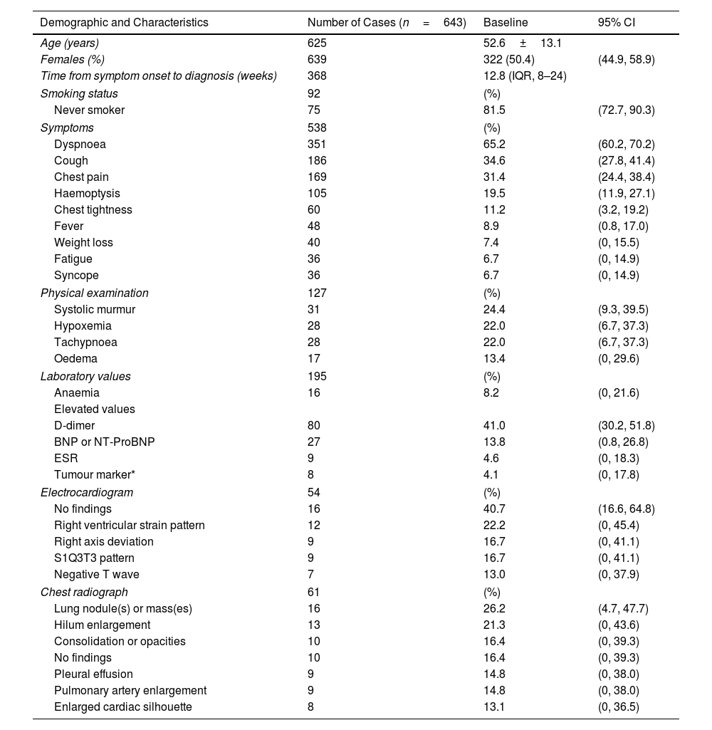
Table 1. Baseline Clinical Characteristics of PPAS.
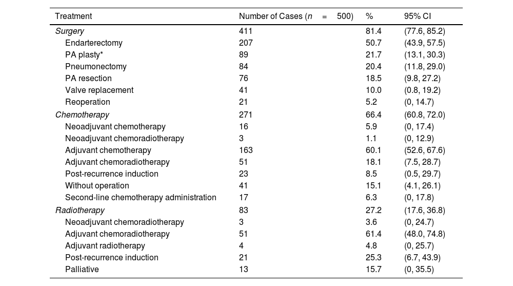
Table 2. Treatment of PPAS.
Show moreShow less
Additional material (1)
Abstract
Objectives

Primary pulmonary artery sarcoma (PPAS) is a rare disease with unclear clinical manifestations. Advances in imaging devices have improved diagnostic capabilities, potentially affecting clinical characteristics and overall survival (OS); however, details remain unclear. This study conducted a pooled analysis of case reports and series to analyse the clinical characteristics and OS of PPAS in the era of advanced medical devices.

Methods

Data were sourced from PubMed and CINAHL, focusing on studies published between 1 January 2014 and 31 December 2023. The study included patients diagnosed with PPAS, with extracted data covering demographics, diagnosis, treatments, and survival.

Results

Overall, 643 patients were included (mean age: 52.6±13.1 years; 50.4% were female). Initially, 70.6% were diagnosed with pulmonary thromboembolism (PTE), and 15.4% were suspected of having PPAS. Among these, 93.9% and 55.2% showed suggestive findings on computed tomography (CT)-integrated positron emission tomography with 2-deoxy-2-18F-fluoro-d-glucose (18F-FDG PET/CT) and CT, respectively, with 98.2% confirmed before death. The right main pulmonary artery was the most affected site on CT (72.3%). Surgery and chemotherapy were performed in 81.4% and 66.4% of patients, respectively. The median OS was 31 months, with surgery extending OS across all stages and chemotherapy benefiting stages III–IV. Longer OS was achieved in patients who underwent complete surgical resection.

Conclusions

18F-FDG PET/CT and multi-detector-row CT can differentiate PTE from PPAS. These medical devices may contribute to improved OS.

Keywords:
Primary pulmonary artery sarcoma
Multi-detector row computed tomography
18F-FDG PET/CT
Pulmonary thromboembolism
Overall survival
Review
Abbreviations:
PPAS
PAIS
MDCT
18F-FDG PET/CT
EBUS-TBNA
ECGF
PRISMA
ROB
NOS
RVOT
IQR
OS
HR
BNP
MRI
SUVmax
PTE
TBB
AI
TNM
MDM2
PDGFRA
EGFR
CDKN2A
Graphical abstract
Full Text
Introduction

Primary pulmonary artery sarcoma (PPAS) is a mesenchymal tumour originating in the pulmonary artery.1,2 Although the incidence of PPAS is unknown, estimates suggest it ranges from 0.001% to 0.03%.3 This rare disease often develops predominantly within the pulmonary artery lumen, thus complicating diagnosis and treatment owing to the absence of early symptoms and delays in detection. Moreover, the pulmonary artery's critical role in blood circulation further complicates its management. Although periodic updates on PPAS have been published,4,5 its clinical features remain unclear, and contemporary treatments reflecting recent medical advancements have not been sufficiently addressed. Consequently, the pathogenesis of PPAS remains poorly understood, with many aspects unexplored, including its origin site, primary lesion characteristics, the distinction between pulmonary artery intimal sarcoma (PAIS) and pulmonary artery luminal sarcoma, optimal treatment strategies, and recurrence patterns.

Advancements in medical technology have provided some insight into PPAS, albeit to a limited extent. Tools including multi-detector-row computed tomography (MDCT),6 CT-integrated positron emission tomography with 2-deoxy-2-18F-fluoro-d-glucose (18F-FDG PET/CT),7 and high-resolution echocardiography,8 which gained global popularity in the 21st century, have significantly improved diagnostic capabilities. Additionally, emerging diagnostic techniques, such as endobronchial ultrasound-guided transbronchial needle aspiration (EBUS-TBNA)9 and endovascular catheter-guided forceps biopsy (ECGF)10 may further advance diagnosis. Recent progress in genetic testing,11–13 immune checkpoint inhibitors,14 and molecular-targeted therapies12 has also been reported.

Mandelstam first reported PPAS in 1923.15 Modern medical technology requires continuous updates to diagnosis and treatment strategies to improve prognosis. Acknowledging the role of current research in advancing medical technology, we conducted a pooled analysis of case reports and series to gather relevant information on PPAS. This study focused on data between 2014 and 2023, aligning with the broad adoption of MDCT and 18F-FDG PET/CT for diagnosis.

Methods

This pooled analysis was conducted and reported following the Preferred Reporting Items for Systematic Reviews and Meta-Analyses (PRISMA) guidelines.16 The study was registered with the University Hospital Medical Information Network Clinical Trials Registration (registration ID number UMIN000053256).17

Search and Extraction

We conducted a search of PubMed and CINAHL for studies published between 1 January 2014 and 31 December 2023. The search terms used were: ‘pulmonary artery’ and ‘sarcoma’ or ‘intimal sarcoma’. The quality assessment of all included studies was conducted by two reviewers (F.K. and H.C.). Studies requiring additional evaluation were reviewed by a third reviewer (N.T.) and resolved through discussions among the three reviewers.

Inclusion and Exclusion Criteria

The inclusion criteria were: (1) confirmed diagnosis of PPAS; (2) availability of baseline clinical characteristics (e.g., age, sex, and symptoms), diagnosis, or treatment data; (3) clinical studies, observational studies, case series, and case reports; and (4) studies in any language. The exclusion criteria were: (1) in vitro or animal experiments, reviews, meta-analyses, and duplicates and (2) conference abstracts.

Definition

PPAS is defined as a neoplasm originating from mesenchymal cells between the right ventricular outflow tract (RVOT) and pulmonary artery. Patients were classified using the staging system for PPAS proposed by Blackmon et al.18: Stage I, tumour limited to the main pulmonary artery; Stage II, tumour involving one lung plus a main pulmonary artery; Stage III, bilateral lung involvement; and Stage IV, extrathoracic spread.

Outcomes

Given the exploratory nature of the pulmonary artery sarcoma data, primary outcomes focused on clinical features, while secondary outcomes involved comparative analyses of overall survival (OS) by treatment in patients with PPAS. Survival was analysed with or without surgery and chemotherapy, categorised into stages I–II and III–IV. Statistical methods are detailed in subsequent sections.

Risk of Bias

The Newcastle–Ottawa Quality Assessment Scale (NOS) and a tool for evaluating the methodological quality of case reports and case series were used to identify the risk of bias.19,20 Owing to the nature of single-arm studies, case series studies (>5) received a maximum of four stars, case–control studies five stars, and cohort studies six stars. Case series (≤5) and case reports were evaluated using the methodological quality tool, with a maximum of five stars. Studies were included if all evaluable items in the NOS and tool received over two stars.

Statistical Analysis

All data were analysed using JMP Pro version 17.1.0 (SAS Institute Inc., Tokyo, Japan) and the Review Manager v. 5.4.1 software (RevMan) (Cochrane Collaboration, Oxford, UK). Continuous variables are expressed as means and standard deviations following a normal distribution; otherwise, the median and interquartile range (IQR) are used. RevMan was used to conduct meta-analysis. For the secondary endpoint of OS, the Kaplan–Meier method was used to estimate the median OS. The stratified log-rank method was used to compare the median OS after surgical and chemotherapeutic interventions. Furthermore, subgroup analyses were performed according to stage and follow-up completion status. Proportional hazards were determined for these OS analyses, and hazard ratios (HRs) with 95% confidence intervals (CIs) were estimated using stratified Cox proportional hazards models, with subgroup analyses presented in forest plots. Statistical significance was set at p<0.05 and was determined using the log-rank test.

ResultsSelection of Eligible Studies

A preliminary search identified 458 relevant studies from two databases (PubMed=387, CINAHL=71). After removing 56 duplicate records, 402 studies were screened based on the titles and abstracts. Among these, 44 were excluded for not meeting the inclusion criteria, resulting in 358 studies for eligibility assessment. Subsequently, 18 studies were excluded for lacking information, 23 for metastatic pulmonary artery sarcoma, 52 for other diseases, and 1 study was withdrawn, as shown in the PRISMA flowchart (Fig. S1).

Included Studies

Finally, 264 studies involving 643 patients were analysed. These included 1 pilot study, 1 preliminary study, 1 cohort study, 4 case–control studies, 35 case series, and 222 case reports. No phase II or III clinical trials were identified (Fig. S1). Twenty-eight studies were evaluated using NOS. One study was evaluated on a six-star scale, five on a five-star scale, and 22 on a four-star scale (Supplementary List 1). All the analysed case reports were judged to be included in the overall appraisal of tools for evaluation of the methodological quality of case reports and case series (Supplementary List 2).20

Primary Outcomes

The clinical features of PPAS are summarised in Table 1. The mean age of patients with PPAS was 52.6±13.1 years. Sex distribution was approximately equal, with 317 male (49.6%) and 322 female (50.4%) patients; 81.5% were non-smokers. Common initial symptoms included dyspnoea (65.2%), cough (34.6%), and chest pain (31.4%). The duration from symptom onset to diagnosis was 12.8 (IQR, 8–24) weeks. Physical examinations showed systolic murmurs (24.4%), hypoxemia (22.0%), tachypnoea (22.0%), and oedema (13.4%). Haematological tests revealed slightly elevated D-dimer levels in 41.0% of patients, elevated brain natriuretic peptide (BNP) or N-terminal pro-BNP levels (13.8%), and anaemia (8.2%). Electrocardiograms were normal in 40.7% of cases, while 38.9% indicated right heart overload. Chest radiographs showed lung nodules or masses (26.2%), hilum enlargement (21.3%), consolidation or opacities (16.4%), pleural effusion (14.8%), pulmonary artery enlargement (14.8%), and an enlarged cardiac silhouette (13.1%); 16.4% showed no abnormalities.

Table 1.

Baseline Clinical Characteristics of PPAS.

Demographic and Characteristics  Number of Cases (n=643)  Baseline  95% CI 
Age (years)  625  52.6±13.1   
Females (%)  639  322 (50.4)  (44.9, 58.9) 
Time from symptom onset to diagnosis (weeks)  368  12.8 (IQR, 8–24)   
Smoking status  92  (%)   
Never smoker  75  81.5  (72.7, 90.3) 
Symptoms  538  (%)   
Dyspnoea  351  65.2  (60.2, 70.2) 
Cough  186  34.6  (27.8, 41.4) 
Chest pain  169  31.4  (24.4, 38.4) 
Haemoptysis  105  19.5  (11.9, 27.1) 
Chest tightness  60  11.2  (3.2, 19.2) 
Fever  48  8.9  (0.8, 17.0) 
Weight loss  40  7.4  (0, 15.5) 
Fatigue  36  6.7  (0, 14.9) 
Syncope  36  6.7  (0, 14.9) 
Physical examination  127  (%)   
Systolic murmur  31  24.4  (9.3, 39.5) 
Hypoxemia  28  22.0  (6.7, 37.3) 
Tachypnoea  28  22.0  (6.7, 37.3) 
Oedema  17  13.4  (0, 29.6) 
Laboratory values  195  (%)   
Anaemia  16  8.2  (0, 21.6) 
Elevated values       
D-dimer  80  41.0  (30.2, 51.8) 
BNP or NT-ProBNP  27  13.8  (0.8, 26.8) 
ESR  4.6  (0, 18.3) 
Tumour marker*  4.1  (0, 17.8) 
Electrocardiogram  54  (%)   
No findings  16  40.7  (16.6, 64.8) 
Right ventricular strain pattern  12  22.2  (0, 45.4) 
Right axis deviation  16.7  (0, 41.1) 
S1Q3T3 pattern  16.7  (0, 41.1) 
Negative T wave  13.0  (0, 37.9) 
Chest radiograph  61  (%)   
Lung nodule(s) or mass(es)  16  26.2  (4.7, 47.7) 
Hilum enlargement  13  21.3  (0, 43.6) 
Consolidation or opacities  10  16.4  (0, 39.3) 
No findings  10  16.4  (0, 39.3) 
Pleural effusion  14.8  (0, 38.0) 
Pulmonary artery enlargement  14.8  (0, 38.0) 
Enlarged cardiac silhouette  13.1  (0, 36.5) 

Abbreviations: CI, confidence interval; IQR, interquartile range; BNP, brain natriuretic peptide; NT-ProBNP, N-terminal fragment brain natriuretic peptides; ESR, erythrocyte sedimentation rate.

*

CA125, NSE, AFP, SCC.

CT revealed abnormalities in the pulmonary artery (dilation, contrast loss, or infiltration) in all the cases. The right main pulmonary artery was the most frequently invaded lesion (72.3%, 95% CI 70.9–79.6), significantly more common than the left main pulmonary artery (58.9%, 95% CI 53.9–63.8; p=0.04) (Fig. 1A and Fig. S2).

Fig. 1.

Distribution of primary lesion of primary pulmonary artery sarcoma. (A) CT lesions at initial examination. In the analysis of 384 cases, lesions were more commonly found in the right main pulmonary artery than in the left, with a significant difference (p=0.04). (B) Presumptive primary lesion of PPAS. In the analysis of 170 cases with confirmed primary lesions, the right main pulmonary artery was the most common site. However, no significant difference was observed (p=0.22). CI, confidence interval.

(0.25MB).

On magnetic resonance imaging (MRI), the most common finding was T2 high heterogeneous lesions (including fat-suppressed hyperintense lesions on T2-weighted imaging), observed in 68.5% of the cases (Table S1). Transthoracic echocardiography frequently showed increased systolic pulmonary artery pressure (46.8%) and, in some cases, mass lesions (18.3%) (Table S2). Using a maximum standardised uptake value (SUVmax) cut-off value of 3.3,21 the positivity rate of 18F-FDG PET/CT was 93.9%, with a median SUVmax of 7.2 (IQR, 5.8–10.3) (Table S3). Pulmonary hypertension, as assessed by right ventricular catheterisation, showed a median mean pulmonary artery pressure of 66.5 (IQR, 44.8–68.0) mmHg (Table S4).

At the time of initial diagnosis, 70.6% of patients were suspected of having pulmonary artery thromboembolism (PTE), whereas 15.4% were suspected of PPAS (Table S5). In instances where PPAS was initially suspected, CT findings contributed to 55.2% of these suspicions. Evaluations using MDCT or enhanced CT were useful in many cases (Table S6).

The distribution of patients according to stage was as follows: Stage I, 34.4%; Stage II, 46.8%; Stage III, 16.0%; and Stage IV, 2.8% (Fig. 2).

Fig. 2.

Clinical stage of primary pulmonary artery sarcoma. Based on the staging by Blackmon et al., 543 patients with PPAS were staged as follows: Stage I (34.4%), Stage II (46.8%), Stage III (16.0%), and Stage IV (2.8%). Stage I: Tumour in the main pulmonary artery; Stage II: Tumour in one lung plus the main artery; Stage III: Bilateral lung involvement; Stage IV: Extrathoracic spread. CI, confidence interval.

(0.09MB).

Surgery was the most frequently performed procedure for a definitive diagnosis, accounting for 72.2% of cases. The most common non-surgical procedure was ECGF, performed in 12.3% of cases (Fig. S3). The confirmed autopsy diagnosis rate was 1.8% in all patients (Fig. S3). The complication rate for diagnostic procedures was 12.5%, with grade 5 complications occurring in 7.9% of the cases (Table S7). Although the number of cases was small, definitive diagnoses were obtained using EBUS-TBNA in 57.1% of cases and transbronchial biopsy in 35.7% of cases (Table S8).

Histopathological analyses included immunohistochemistry (Table S9), genetic testing (Table S10), and subtype classifications obtained from surgery and autopsy (Fig. S4). Additional analyses were performed on the specimens obtained from all procedures (Fig. S5) and biopsy samples (Fig. S6). Common histopathological subtypes identified during surgery and autopsy were undifferentiated sarcomas (23.9%), pleomorphic sarcomas (12.1%), and myxofibrosarcomas (11.8%). The frequencies of these subtypes vary according to the diagnostic techniques used. The primary lesion was histopathologically identified in some surgical and autopsy cases. The right main pulmonary artery was the most frequently identified primary lesion (35.8%), with no significant differences (Fig. 1B and Fig. S7). PAIS was diagnosed in 80.5% of patients with PPAS.

PPAS was treated surgically in 81.4% of the cases. Among these, the most common procedure was endarterectomy, performed in 50.7% of cases. Other surgical procedures included pulmonary artery plasty with partial wall removal and reconstruction (21.7%) and pneumonectomy (20.4%). Mortality within the first postoperative month was 5.9%, and reoperation was necessary in 5.2% of cases. Chemotherapy was administered to 66.4% of patients; 60.1% received it adjuvantly, 15.1% without surgery, and 6.3% as second-line treatment. The most common regimens were doxorubicin hydrochloride (adriamycin) and ifosfamide (Table S11). Radiation therapy was administered to 27.2% of patients, mostly as adjuvant chemoradiotherapy (61.4%) (Table 2).

Table 2.

Treatment of PPAS.

Treatment  Number of Cases (n=500)  95% CI 
Surgery  411  81.4  (77.6, 85.2) 
Endarterectomy  207  50.7  (43.9, 57.5) 
PA plasty*  89  21.7  (13.1, 30.3) 
Pneumonectomy  84  20.4  (11.8, 29.0) 
PA resection  76  18.5  (9.8, 27.2) 
Valve replacement  41  10.0  (0.8, 19.2) 
Reoperation  21  5.2  (0, 14.7) 
Chemotherapy  271  66.4  (60.8, 72.0) 
Neoadjuvant chemotherapy  16  5.9  (0, 17.4) 
Neoadjuvant chemoradiotherapy  1.1  (0, 12.9) 
Adjuvant chemotherapy  163  60.1  (52.6, 67.6) 
Adjuvant chemoradiotherapy  51  18.1  (7.5, 28.7) 
Post-recurrence induction  23  8.5  (0.5, 29.7) 
Without operation  41  15.1  (4.1, 26.1) 
Second-line chemotherapy administration  17  6.3  (0, 17.8) 
Radiotherapy  83  27.2  (17.6, 36.8) 
Neoadjuvant chemoradiotherapy  3.6  (0, 24.7) 
Adjuvant chemoradiotherapy  51  61.4  (48.0, 74.8) 
Adjuvant radiotherapy  4.8  (0, 25.7) 
Post-recurrence induction  21  25.3  (6.7, 43.9) 
Palliative  13  15.7  (0, 35.5) 

Abbreviations: CI, confidence interval; PA, pulmonary artery.

*

Part of the wall was removed and reconstructed.

The primary lesion area was the most frequent recurrence site in patients with PPAS (42.5%). Other common recurrence sites included the bone (11.3%), brain (8.5%), and liver (6.6%) (Table S12).

The pooled analysis conducted using RevMan yielded similar results, revealing comparable gender ratios and proportions of patients with dyspnoea, cough, PH comorbidity, and those receiving adjuvant chemotherapy. However, differences were noted in the proportions of patients with chest pain and those diagnosed with PTE or PPAS at the initial diagnosis (Fig. S8).

Secondary Outcomes

The secondary endpoint explored the effects of surgical and chemotherapeutic interventions on OS among patients with PPAS, which was 31 months overall (Fig. 3).

Fig. 3.

Overall survival in patients with primary pulmonary artery sarcoma. Median OS of all patients with PPAS was 31 months. PPAS, primary pulmonary artery sarcoma; OS, overall survival.

(0.11MB).

Both interventions improved OS (Fig. 4).

Fig. 4.

Overall survival (OS) with or without intervention in patients with all-stage primary pulmonary artery sarcoma. (A) Surgical intervention. (B) Chemotherapeutic intervention. Surgery and chemotherapy improved OS in patients with all-stage PPAS. PPAS, primary pulmonary artery sarcoma; OS, overall survival; CI, confidence interval.

(0.25MB).

Subgroup analysis revealed that surgery enhanced OS in stages I–II (Fig. S9), whereas chemotherapy showed no significant difference (Fig. S10). In stages III–IV, both interventions extended OS (Figs. S11 and S12). However, surgery with adjuvant chemotherapy improved the prognosis of Stage I–II (Figs. S13, S14, and S15). The forest plot showed a 95% CI below 1 for all treatments, except for chemotherapy in stages I–II (Fig. S16). Furthermore, there was no difference in OS between patients with PPAS who underwent pneumonectomy and those who underwent other surgical procedures (Fig. S17). However, regardless of the procedure, there was a significant difference in median OS when comparing patients with PPAS who underwent complete resection to those who did not (p<0.001) (Fig. S18).

Discussion

Our findings showed no significant sex differences, with most being non-smokers. The risk factors for PPAS remain unclear. However, a patient with PPAS having osteosarcoma implicated vinyl chloride exposure as a pathogenic factor.22 Additionally, dioxides, arsenic, anabolic steroids, and foreign bodies were considered risk factors for angiosarcoma.23 External environmental factors seem to impact the development of PPAS. The symptoms and physical findings were nonspecific, consistent with previous research. The diagnosis process lasted approximately 12.8 weeks from the onset of symptoms (IQR, 8–24 weeks). Electrocardiograms were normal in 40.7% of cases, and chest radiographs were unremarkable in 16.4% of instances. These delays were primarily due to difficulties in differentiating PPAS from PTE or chronic thromboembolic pulmonary hypertension.24

With advancements in MDCT,6 lesions suggestive of PPAS were observed in 55.2% of cases. Indicators such as the wall-eclipsing sign,6 polylobulated22 and bulging margins proximal to the defect,6,25 and spherical shapes of defects25,26 are distinctive from thrombus-only lesions, suggesting PPAS. Echocardiographic findings, such as the “sieve sign” have also improved the diagnosis of PPAS.8 Likewise, MRI improved the diagnosis.27 The most definitive imaging test was 18F-FDG PET-CT.21 Additionally, although the deficient areas on contrast-enhanced CT were previously thought to be completely occluded by the tumour, 18F-FDG PET-CT confirmed that some areas showed a mixture of uptake and non-uptake regions.7 This finding aids in understanding the pathophysiology. PPAS in the pulmonary artery causes blood flow stasis, vascular endothelial damage, and increased coagulability – known as Virchow's triad – which can result in secondary PTE.28 We highlighted the difficulty in distinguishing PPAS from PTE, with recent autopsy reports suggesting possible coexistence between PPAS and PTE.29 Not all PPAS lesions exhibit characteristic uptake patterns.30 Lesions lacking uptake often display low cell counts and abundant myxoid tissue, compounded by intratumoral haemorrhage, necrosis, mucous,30 or calcification.22 Additionally, 18F-FDG PET-CT uptake is occasionally observed in patients with PTE.31 This uptake may be attributed to the involvement of inflammatory cells, such as macrophages and neutrophils, in the thrombus and organising lesions.32

Our study also addressed the challenges in staging PPAS. OS varied significantly by stage, especially in Stage II, and did not directly correlate with prognosis (data not shown). This variability could be due to comorbidities and inconsistent interventional therapies, underscoring the importance of identifying which patients with PPAS benefit most from chemotherapy.

Furthermore, there are numerous challenges in achieving a definitive diagnosis. While new diagnostic techniques such as ECGF and EBUS-TBNA have been introduced,9,10 there are significant individual differences in diagnostic rates,10 and grade 5 adverse events due to diagnostic procedures were observed in 7.9% of all cases. Additionally, small biopsy specimens were more likely to be diagnosed as undifferentiated sarcoma or spindle cell sarcoma. Furthermore, even when surgery provides sufficient lesions for diagnosis, several subtypes of PPAS may coexist, and the delineation of the tumour remains unclear.25 Conversely, genetic analysis of histopathological specimens has become more prevalent.11,12 Recent findings indicate a positive MDM2 gene in many PAIS cases,13 highlighting its role in tumour progression and genetic factors. Additionally, genes such as PDGFRA,12EGFR, and CDKN2A13 have also been studied.11,33 Notably, the percentage of cases reaching a definitive diagnosis via autopsy decreased significantly, from 59% in a report from the 1980s34 to 5.0% in a report spanning 1990 to 2010.4 Our study revealed a further reduction to 1.8%, underscoring advancements in testing and treatment.

Previous studies reported that PPAS occurs most frequently in the posterior wall of the main pulmonary artery.2 Based on this, we also analysed the primary site of PPAS. While a higher number of patients originated from the right main pulmonary artery, no statistically significant difference was observed. However, analysis of lesion sites on chest CT revealed that the right main pulmonary artery was significantly more frequently affected. The reason for this remains unclear, but it may be influenced by the larger number of patients analysed using CT findings and the anatomical fact that the right main pulmonary artery is longer than the left.35

Several challenges surround the treatment of PPAS: surgery has been the most promising approach for prolonging OS.36 Regardless of the procedure, our study found a significant difference in OS when comparing patients with complete resection or not (p<0.001). One reason is that patients with Stage I of complete resection require only pulmonary artery resection and reconstruction, without the need for pneumonectomy. Thus, factors such as tumour location, size, and metastasis lesion significantly influence outcomes, preventing the establishment of standardised treatment protocols. In our study, we were unable to compare each surgical procedure due to the varied approaches employed. Many patients undergo a combination of surgical procedures or two-stage surgeries, and their lesions and stages widely vary. Based on the results of a retrospective analysis from 1997 to 2010, the mortality rate within 30 days after surgery was reported to be 13%.37 In this study, the mortality rate within 30 days after surgery was 5.9%, suggesting technical improvements in surgery, including perioperative management, although no standard treatment exists. Furthermore, although chemotherapy has historically shown limited efficacy, our study indicates it may extend OS in advanced stages III–IV but not significantly in early stages I–II. This enhancement likely results from selecting effective regimens and integrating multidisciplinary treatments.12,14,38 Limited reports suggest pembrolizumab and anlotinib are effective.33,39 Analysing tumour immunity and the microenvironment in PPAS may identify groups with better prognoses.40 Finally, differences between the two evaluation methods, JMP and RevMan, could be attributed to the greater proportion of studies in the RevMan analysis that focused on surgical patients initially suspected of having PPAS and excluded from PTE.

This study had some limitations. First, it was retrospective. Second, there was publication bias. Third, the analysed patients exhibited various data deficits due to the rarity, diagnostic challenges, and fatal nature of PPAS, leading to the selection of patients with positive results, characteristic of publication bias. Additionally, the study largely comprised case reports and series from databases without clinical trials, complicating systematic analysis. The secondary endpoint, OS by treatment intervention, was also skewed by publication bias, as only patients eligible for surgery, chemotherapy, or radiation therapy were selected. Finally, treatments were analysed as single categories, though specific techniques, regimens, or devices could have influenced OS.

Conclusion

PPAS, an extremely rare tumour historically diagnosed posthumously in the 20th century, currently benefits from pre-mortem diagnoses using modern technologies. Advances in genetic testing and data collection are expected to significantly improve PPAS prognoses.

CRediT Authorship Contribution Statement

F.K. interpreted the data and drafted the original and revised manuscript. H.C. significantly contributed to data analysis and interpretation. N.T. and Y.K. contributed to data analysis and data curation. S.M., R.O., A.M., M.K., C.Y., A.K., K.Y., N.H., H.O., and M.M. were responsible for data curation. H.K., M.M., K.T., and T.K. made substantial contributions to revising the manuscript drafts. All authors have reviewed and approved the final version of the manuscript and agree to be accountable for their respective contributions to the work.

Patient Consent

Not required due to the nature of the pooled analysis.

Institutional Review Board (IRB) approval was exempted due to the nature of the meta-analysis.

The University Hospital Medical Information Network Clinical Trials Registration ID Number

The study was registered with the University Hospital Medical Information Network Clinical Trials Registration (registration ID number UMIN000053256).

Guarantor

Fumihiro Kashizaki

Conflict of Interests

The authors state that they have no conflict of interests.

Acknowledgments

We thank all patients with primary pulmonary artery sarcoma.

Appendix B
Supplementary Data

The following are the supplementary data to this article:

References
[1]
C.D.M. Fletcher, K. Unni, F. Mertens.
WHO classification of tumours of soft tissue and bone.
5th ed., IARC Press, (2020),
[2]
A.P. Burke, R. Virmani.
Sarcomas of the great vessels. A clinicopathologic study.
[3]
E.Y. Choi, Y.W. Yoon, H.M. Kwon, D. Kim, B.E. Park, Y.S. Hong, et al.
A case of pulmonary artery intimal sarcoma diagnosed with multislice CT scan with 3D reconstruction.
Yonsei Med J, 45 (2004), pp. 547-551
[4]
D. Bandyopadhyay, T.S. Panchabhai, N.S. Bajaj, P.D. Patil, M.C. Bunte.
Primary pulmonary artery sarcoma: a close associate of pulmonary embolism-20-year observational analysis.
J Thorac Dis, 8 (2016), pp. 2592-2601
[5]
T. Assi, J. Kattan, E. Rassy, T. Moussa, H. Nassereddine, C. Honore, et al.
A comprehensive review on the diagnosis and management of intimal sarcoma of the pulmonary artery.
Crit Rev Oncol Hematol, 147 (2020), pp. 102889
[6]
H.L. Gan, J.Q. Zhang, X.Y. Huang, W. Yu.
The wall eclipsing sign on pulmonary artery computed tomography angiography is pathognomonic for pulmonary artery sarcoma.
[7]
B. Pan, S.C. Wang, Z.K. Chen, X. Chen.
Primary pulmonary artery sarcoma with intrapulmonary metastases based on PET/CT imaging: a case report and literature review.
Ann Palliat Med, 10 (2021), pp. 7013-7018
[8]
W. Jiang, M. Liu, X. Guo, J. Li, J. Gong, M. Yang, et al.
Echocardiographic characteristics of pulmonary artery intimal sarcoma: comparison with CTPA.
Heart Lung Circ, 32 (2023), pp. 1080-1088
[9]
T. Villeneuve, S. Pontier, G. Prevot, N. Guibert, C. Chevreau, P. Rochaix, et al.
EBUS-TBNA for the diagnosis of primary pulmonary artery sarcoma.
Respir Med Res, 83 (2023), pp. 100974
[10]
J.K. Yeh, S.Y. Hsiao, J.N. Roan, Y.P. Chen, P.Y. Liu, H.Y. Chang.
Endovascular biopsy for pulmonary artery sarcoma mimicking pulmonary embolism.
Acta Cardiol Sin, 39 (2023), pp. 658-662
[11]
C. Koelsche, J.K. Benhamida, F.K.F. Kommoss, D. Stichel, D.T. Jones, S.M. Pfister, et al.
Intimal sarcomas and undifferentiated cardiac sarcomas carry mutually exclusive MDM2, MDM4, and CDK6 amplifications and share a common DNA methylation signature.
Mod Pathol, 34 (2021), pp. 2122-2129
[12]
S. Sai, Y. Imamura, N. Kiyota, N. Jimbo, M. Toyoda, Y. Funakoshi, et al.
Relationship between PDGFR expression and the response to pazopanib in intimal sarcoma of the pulmonary artery: a case report.
Mol Clin Oncol, 14 (2021), pp. 6
[13]
C.E. Wu, C.T. Ng, K.T. Tan.
Transient response of olaparib on pulmonary artery sarcoma harboring multiple homologous recombinant repair gene alterations.
[14]
J.E. Birkness-Gartman, D.L. Thomas, L.L. Engle, L. Voltaggio, E.D. Thompson.
Immune microenvironment of intimal sarcomas: adaptive immune resistance with potential therapeutic implications.
Am J Clin Pathol, 161 (2024), pp. 256-263
[15]
M. Mandelstamm.
Uber primare neubildungen des herzens.
Virchows Arch Pathol Anat, 245 (1923), pp. 43-54
[16]
M.J. Page, J.E. McKenzie, P.M. Bossuyt, I. Boutron, T. Hoffmann, C.D. Mulrow, et al.
Mapping of reporting guidance for systematic reviews and meta-analyses generated a comprehensive item bank for future reporting guidelines.
J Clin Epidemiol, 118 (2020), pp. 60-68
[17]
University Hospital Medical Information Network [accessed 21.06.24]. Available from: https://center6.umin.ac.jp/cgi-bin/ctr_e/ctr_view.cgi?recptno=R000060777
[18]
S.H. Blackmon, D.C. Rice, A.M. Correa, R. Mehran, J.B. Putnam, W.R. Smythe, et al.
Management of primary pulmonary artery sarcomas.
Ann Thorac Surg, 87 (2009), pp. 977-984
[19]
A. Stang.
Critical evaluation of the Newcastle–Ottawa scale for the assessment of the quality of nonrandomized studies in meta-analyses.
Eur J Epidemiol, 25 (2010), pp. 603-605
[20]
M.H. Murad, S. Sultan, S. Haffar, F. Bazerbachi.
Methodological quality and synthesis of case series and case reports.
BMJ Evid Based Med, 23 (2018), pp. 60-63
[21]
X.Y. Xi, W. Gao, J.N. Gong.
Value of (18)F-FDG PET/CT in differentiating malignancy of pulmonary artery from pulmonary thromboembolism: a cohort study and literature review.
Int J Cardiovasc Imaging, 35 (2019), pp. 1395-1403
[22]
Y. Zheng, X. Duan, H. Wang, S. Zhao.
Pulmonary artery osteosarcoma masquerading as pulmonary thromboembolism: the role of multimodality imaging.
ESC Heart Fail, 8 (2021), pp. 5565-5567
[23]
R.J. Young, N.J. Brown, M.W. Reed, D. Hughes, P.J. Woll.
Angiosarcoma.
Lancet Oncol, 11 (2010), pp. 983-991
[24]
F. Ampatzidou, O. Drosos, A. Madesis, G. Drossos.
Pulmonary artery sarcoma presenting as chronic thromboembolic pulmonary hypertension (CTEPH).
Kardiochir Torakochirurgia Pol, 20 (2023), pp. 129-131
[25]
M. Monteagudo-Vela, A. Rosell Alayza, E. Monguió Santín, A. Cecconi, G. Reyes Copa.
Pulmonary artery sarcoma with extensive leiomyosarcoma differentiation and heterologous osteosarcomatous elements.
Cardiovasc Pathol, 60 (2022), pp. 107436
[26]
K. Freystaetter, A. Ismailbaev, A. Simonyan, S. Chernyavskiy, A. Goodwin, R. Komarov.
Radical surgery for recurrent pulmonary artery sarcoma with 4-year survival.
Ann Thorac Surg, 113 (2022), pp. e331-e334
[27]
M.X. Liu, Z.H. Ma, T. Jiang.
Differential diagnosis of pulmonary artery sarcoma and central chronic pulmonary thromboembolism using CT and MR images.
Heart Lung Circ, 27 (2018), pp. 819-827
[28]
A. Labianca, T. Bosetti, A. Indini, G. Negrini, R.F. Labianca.
Risk prediction and new prophylaxis strategies for thromboembolism in cancer.
Cancers (Basel), 12 (2020),
[29]
A. Ro, S. Mori, H. Sugiura.
An autopsy case of pulmonary artery intimal sarcoma: detailed observation of tumor and its related lesions in pulmonary arteries.
Cardiovasc Pathol, 43 (2019), pp. 107143
[30]
H. Suto, M. Suto, Y. Inui, A. Okamura.
Difficulty in distinguishing pulmonary arterial intimal sarcoma from pulmonary thromboembolism using FDG PET/CT.
In Vivo, 36 (2022), pp. 1519-1522
[31]
R.R. Flavell, S.C. Behr, R.L. Brunsing, D.M. Naeger, M.H. Pampaloni.
The incidence of pulmonary embolism and associated FDG-PET findings in IV contrast-enhanced PET/CT.
Acad Radiol, 21 (2014), pp. 718-725
[32]
C. Wittram, J.A. Scott.
18F-FDG PET of pulmonary embolism.
AJR Am J Roentgenol, 189 (2007), pp. 171-176
[33]
Y. Mounai, T. Yoshida, S. Ito.
Pulmonary artery intimal sarcoma in a patient with lynch syndrome: response to an immune checkpoint Inhibitor.
Case Rep Oncol, 16 (2023), pp. 21-29
[34]
H.K. Lyerly, J.G. Reves, D.C. Sabiston Jr..
Management of primary sarcomas of the pulmonary artery and reperfusion intrabronchial hemorrhage.
Surg Gynecol Obstet, 163 (1986), pp. 291-301
[35]
M.B. Boumpouli, S.M. Kazakidi Asimina.
Computational analysis of blood flow in healthy pulmonary arteries in comparison to repaired tetralogy of fallot results: a small cohort study.
[36]
J.M. Parish, E.C. Rosenow 3rd, S.J. Swensen, T.B. Crotty.
Pulmonary artery sarcoma. Clinical features.
Chest, 110 (1996), pp. 1480-1488
[37]
S. Mussot, M.-R. Ghigna, O. Mercier.
Retrospective institutional study of 31 patients treated for pulmonary artery sarcoma.
Eur J Cardiothorac Surg, 43 (2013), pp. 787-793
[38]
T. Sun, G.L. Lu, L.C. Ma, J.Z. Huang, S.B. Xie.
Multimodal echocardiography in the diagnosis of masses localized to the proximal portions of pulmonary arteries.
Int J Cardiovasc Imaging, 38 (2022), pp. 149-158
[39]
Y. Wu, J. Huang, Q. Wang.
Whole-exome sequencing insights into pulmonary artery sarcoma mimicking pulmonary embolism: a case report and review.
Onco Targets Ther, 12 (2019), pp. 6227-6235
[40]
S. Wang, S. Guo, J. Guo, Q. Du, C. Wu, Y. Wu, et al.
Cell death pathways: molecular mechanisms and therapeutic targets for cancer.
MedComm (2020), 5 (2024), pp. e693
Copyright © 2024. SEPAR
Archivos de Bronconeumología
Article options
Tools

Are you a health professional able to prescribe or dispense drugs?