


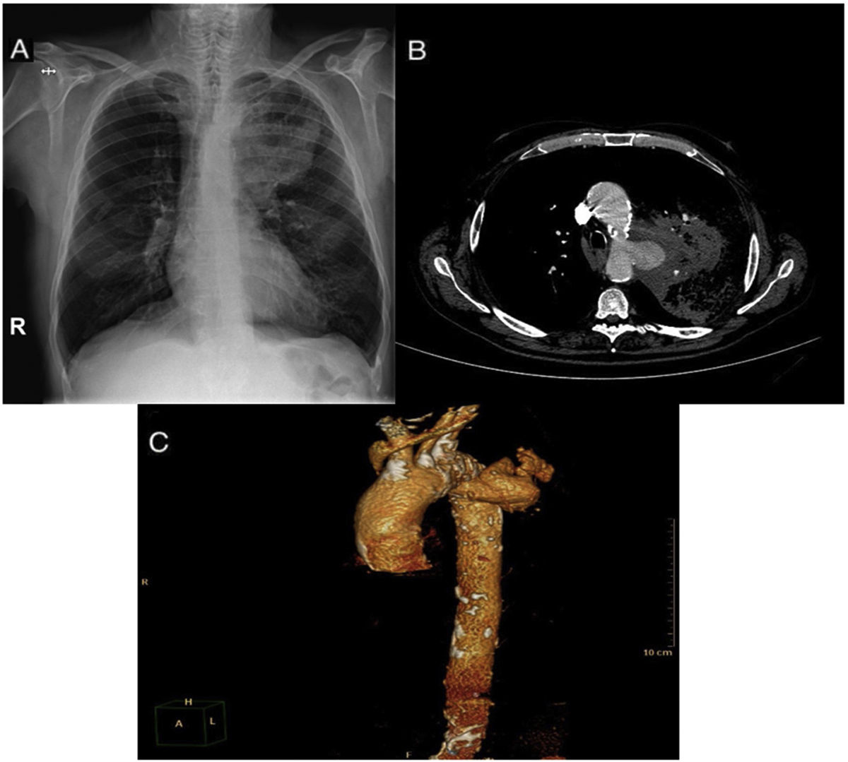
We report the case of a 66-year-old man with hypertension and poorly controlled diabetes who attended the emergency department for moderate hemoptysis (50ml), fever, persistent oppressive central chest pain radiating to the neck, and dyspnea at rest that had developed a few hours previously. He reported a 7-day history of chest pain and blood pressure of up to 200/120mmHg, along with dysphagia to solids in the last 72h. The chest X-ray showed extensive consolidation in the left upper lobe (LUL) with tracheal deviation toward the contralateral side. Emergency chest CT angiogram was requested, revealing a pseudoaneurysm of the aortic arch measuring approximately 4cm×3.5cm, with a 2cm tear in the left lateral aortic wall, an adjacent hematoma, and alveolar hemorrhage in the LUL (Fig. 1).
. Chest CT angiogram imaging showing ruptured pseudoaneurysm with alveolar hemorrhage in LUL (B). Reconstructed 3-dimensional image showing thoracic aorta with ruptured pseudoaneurysm (C).")
Emergency surgery was performed to place an aortic endoprosthesis, but the patient died in the recovery room due to refractory hypovolemic shock associated with left hemothorax.
Thoracic aortic aneurysms are usually asymptomatic, although rupture may constitute a life-threatening emergency.1,2 In our case, onset occurred with hemoptysis and alveolar hemorrhage, and the patient was diagnosed using CT angiogram, although he subsequently died.
The following are the supplementary data to this article:


