Journal Information
Share
Share
Download PDF
More article options
Visits
57
Original Article
Full text access
Uncorrected Proof. Available online 16 July 2024
Apnea-Specific Pulse-Rate Response is Associated With Early Subclinical Atherosclerosis in Obstructive Sleep Apnea
Visits
57
Ana Sanchez-Azofraa,b,
Corresponding author
asanchezazofra@health.ucsd.edu

Corresponding author.
, Jeremy E. Orra, David Sanz-Rubioc, Marta Marin-Otoc,d, Sergio Alarcon-Sisamonc, Eugenio Vicentec, Julio Ancocheab,e, Joan B. Sorianob,e, Pamela DeYounga, Ali Azarbarzinf, Atul Malhotraa, Jose M. Marinc,d,e
a Division of Pulmonary, Critical Care, and Sleep Medicine and Physiology, Department of Medicine, University of California San Diego, 9500 Gilman Dr, La Jolla, CA 92093, USA
b Division of Pulmonary Medicine, Hospital Universitario de la Princesa, Universidad Autónoma de Madrid, Calle Diego de Leon 62, 28006 Madrid, Spain
c Translational Research Unit, IIS Aragón, Hospital Universitario Miguel Servet, Avda. San Juan Bosco 13, 50009 Zaragoza, Spain
d Department of Medicine, Faculty of Medicine, University of Zaragoza, Edificio A, 50009 Zaragoza, Spain
e Center for Biomedical Research on Respiratory Diseases (CIBERES), Instituto de Salud Carlos III, Av. Monforte de Lemos, 3-5, Pabellón 11, Planta 0, 28029 Madrid, Spain
f Division of Sleep and Circadian Disorders, Brigham and Women's Hospital and Harvard Medical School, Harvard University, 75 Francis St, 02115 Boston, MA, USA
This item has received
Article information
Abstract
Full Text
Bibliography
Download PDF
Statistics
Figures (4)
Show moreShow less
Tables (3)
Table 1. Baseline characteristics of study participants.b
Table 2. Regression summary statistics for association between delta pulse rate and CIMT/atheroma plaques.a
Table 3. Regression summary statistics for association between hypoxic burden and T90 with CIMT/atheroma plaques.a
Show moreShow less
Additional material (1)
Abstract
Introduction

In patients with obstructive sleep apnea (OSA), novel metrics such as hypoxic burden (HB) and sleep apnea-specific pulse-rate response (ΔHR) may better correlate with cardiovascular diseases (CVD) than the apnea–hypopnea index (AHI). This manuscript aims to assess the correlation between ΔHR and HB with subclinical atherosclerosis in patients with OSA, testing the hypothesis that elevated ΔHR and HB are associated with subclinical atherosclerosis development.

Methods

In a prospective study, individuals aged 20–65 years with suspected OSA without known comorbidities were consecutively recruited and defined as OSA (AHI5events/h) or healthy controls. Using bilateral carotid ultrasonography, common carotid intima-media thickness (CIMT) was assessed and the identification of at least one atheromatous plaque defined the presence of subclinical atherosclerosis. ΔHR, and HB were derived from pulse-oximetry.
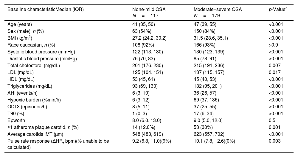
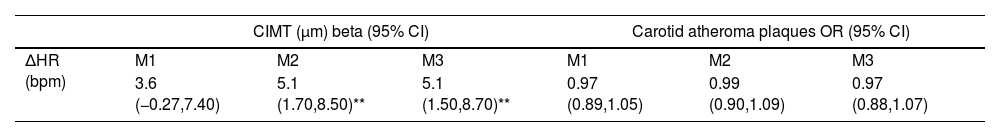
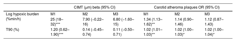
Results

We studied 296 patients of 45±10 years old, of whom 28% were women, and a BMI of 30.3±5.3kg/m2. Overall, 245 had OSA and 51 were healthy controls. After controlling for confounding variables higher ΔHR but not HB, was associated with higher CIMT (p=0.006) and higher time spent with oxygen saturation below 90% (T90) was associated with an increase in carotid atheroma plaques (p=0.032). When stratifying OSA based on HB tertiles, we observed that within tertile 2 of HB, an increase in ΔHR was associated with larger CIMT (p=0.017).

Conclusion

A higher ΔHR is associated with an increase in CIMT among adult patients with OSA. This study suggests that ΔHR could be a biomarker of risk for CVD in patients with OSA.

Keywords:
OSA
Cardiovascular risk
Carotid intima-media thickness
Atheroma plaques
Hypoxic burden
Intermittent hypoxia
Abbreviations:
AHI
BMI
CIMT
CPAP
CVD
EPIOSA
ΔHR
ESS
HB
MESA
M1
M2
M3
ODI
OSA
T90
Full Text
Introduction

Obstructive sleep apnea (OSA) represent the most prevalent form of sleep-disordered breathing, with nearly 1 billion people affected worldwide.1 Despite its widespread prevalence, OSA remains largely undiagnosed.1 In 2019, there were 523 million people around the world affected by cardiovascular diseases (CVD), which accounted for 18.6 million deaths worldwide – close to a third of all deaths globally. OSA is a risk factor for CVD, contributing to conditions such as systemic hypertension, stroke, heart failure, atrial fibrillation, and coronary heart disease.2 The impact of OSA on CVD seems to vary according to severity, with severe OSA consistently associated with increased risks of all-cause and cardiovascular mortality.3,4 Continuous positive airway pressure (CPAP) is the most common and effective treatment for OSA. Nevertheless, whether this treatment is beneficial in reducing cardiovascular risk remains unclear.5–10 New biomarkers that help identify patients at higher risk of developing cardiovascular disease could help clarify which individuals would benefit the most from CPAP treatment.

As our understanding of the physiological features of sleep disordered-breathing has advanced, it has become apparent that the apnea–hypopnea index (AHI), the metric used to assess the severity of OSA by primarily focusing on the frequency of respiratory events, is not an ideal prognostic biomarker for OSA and may not comprehensively capture the entirety of the condition.3,4,11 Despite its widespread acceptance and convenience, the AHI possesses inherent shortcomings that oversimplify the intricate nature of OSA by overlooking various factors contributing to sleep disruption, including the duration of oxygen desaturation, the degree of arousal from sleep, the patterns of respiratory events, and the activation of the autonomic system.11 Consequently, these limitations result in a weak correlation between AHI and the consequences of OSA.

Polysomnography, the gold standard diagnostic procedure of sleep apnea, offer valuable insights into the sleeping, respiratory, and cardiovascular status of patients. As a result, there have been suggestions for alternative metrics to gauge the severity of OSA and better capture CVD risk, relying on advanced signal processing and other sophisticated analyses, including (1) hypoxic burden, measuring the integrated time and depth of intermittent desaturations, and (2) sleep apnea-specific pulse-rate response (ΔHR), reflecting the cortical arousals and autonomic system responsiveness.12–14 Previously, Azarbarzin et al. described a U-shaped relationship between sleep apnea-specific ΔHR and cardiovascular risk scores, as well as an association between higher hypoxic burden and elevated morbidity and mortality risks.15,16

Atherosclerosis is the primary intermediate mechanism explaining most cardiovascular events.17 Previous studies showed that OSA can lead to early atherosclerosis, marked by increased carotid intima-media thickness (CIMT) and atheroma plaque formation, even without significant comorbidities.18–20 The underlying mechanisms behind this association include inflammation, oxidative stress, the autonomic nervous system, vascular dysfunction, platelet activation, metabolic dysfunction, small molecule RNA, and cardioprotective functions.21 We previously showed that plasma levels of oxLDL in OSA patients synergistically contribute with intermittent hypoxia to activate the NLRP3 inflammasome, leading to the subsequent release of tissue factor and the development of early subclinical atherosclerosis.22 Additionally, given that CVD evolves over decades, diagnosing atherosclerosis at a subclinical stage in younger, presumably healthy populations who might benefit from prevention is essential.

Based on this conceptual framework, we tested the hypothesis that high sleep apnea-specific ΔHR and hypoxic burden are associated with the development of subclinical atherosclerosis, measured by CIMT and atheroma plaques, in OSA individuals.

Methods

The Epigenetics Modification in Obstructive Sleep Apnea (EPIOSA) study (ClinicalTrials.gov identifier: NCT02131610) is an ongoing prospective cohort study designed to evaluate inflammatory and epigenetic mechanisms involved in CVD complication patients with OSA.25 Participants aged 20–65 years free of clinically significant comorbid conditions were consecutively recruited at the Sleep Clinic of the Hospital Universitario Miguel Servet, Zaragoza, Spain (Fig. 1). The full inclusion/exclusion criteria are shown in Table S1 in the Supporting Information. The Institutional Review Board at Aragón Research Institute, Human Research Protection Program (03/2013), approved the EPIOSA Study protocol, and all participants provided written informed consent before being enrolled. The present study is a cross-sectional analysis of the EPIOSA cohort. This research followed and endorsed the STROBE guidance for reporting observational research.26 Our manuscript adheres to the ethical guidelines outlined in the Helsinki Declaration, ensuring the protection of human subjects and the integrity of our research.

Fig. 1.

Flow diagram presenting ascertainment of the study sample. ΔHR: sleep apnea-specific pulse-rate response, OSA: obstructive sleep apnea, AHI: apnea–hypopnea index.

(0.15MB).
Measurements

A comprehensive questionnaire was designed for data collection. These dimensions encompassed socio-demographic information, body measurements such as weight, height, waist, hip, and neck circumferences, as well as lifestyle factors like healthy behaviors, daily exercise routines, smoking history, alcohol consumption, and other potentially toxic habits. Additionally, participants’ medical history was collected, including previous diagnoses, comorbidities, personal and family disease history, and current medication use. Daytime somnolence was assessed using the Epworth Sleepiness Scale (ESS).27

A type 3 portable sleep monitor was employed to perform sleep apnea test at home (ApneaLink Air, ResMed, San Diego, CA). The results of all sleep studies were analyzed by trained personnel using standard criteria (AASM Alternative Criteria), and these technicians were blinded to the current protocol.28

Imaging of subclinical atherosclerosis

The CIMT and the presence of atheroma plaque in carotid arteries were determined using the Philips IU22 ultrasound system (Philips Healthcare, Bothell, Washington). The acquisition of ultrasound images was performed through linear high-frequency 2-dimensional probes (Philips Trans-ducer L9-3, Philips Healthcare).29 Carotid plaque was defined as a focal structure protruding ≥0.5mm into the lumen or reaching a thickness of at least ≥50% of the surrounding intima. To quantify CIMT, three measurements were taken on the far wall of both the left and right common carotid arteries in end-diastole, precisely 1cm below the point of bifurcation. The CIMT value was determined by calculating the average of the six readings obtained from these measurements.

Sleep apnea metrics

Classic OSA metrics: An apnea is identified as a cessation of airflow lasting 10s or longer, while a hypopnea is characterized by a reduction in airflow (>50%) lasting at least 10s, accompanied by a decrease in oxygen saturation over 4%. Time spent with oxygen saturation below 90% (T90) was defined as the proportion of T90 recording time with oxygen saturation below 90% in total sleep time.

Sleep apnea-specific pulse-rate response (ΔHR): The pulse rate was obtained from the pulse-oximetry nocturnal signal generated by the Apnealink device and employed to estimate the heart rate. The pulse-oxymetry signal offers a time-averaged heart rate measurement, effectively filtering out beat-to-beat and breath-to-breath variations. This method conveniently enables the evaluation of peak changes in heart rate associated with specific events, which typically occur within a 10–30-s timeframe. In accordance with definitions used in previous studies,12,16 the ΔHR was defined as the difference between a maximum pulse rate within a subject-specific search window and an event-related minimum pulse rate. Finally, the individual-level ΔHR was defined as the mean of all event-specific responses.

Hypoxic burden (HB): To quantify sleep apnea-related hypoxic burden, aimed at capturing the total amount of respiratory event-related hypoxemia over the sleep recording period, we defined the hypoxic burden as the total area under the respiratory event-related desaturation curve, as previously described.15 Briefly, for each individually identified apnea or hypopnea (regardless of associated desaturation), the pre-event baseline saturation was defined as the highest SpO2 level within the 100s leading up to the end of the event. The area under this baseline value was calculated over a subject-specific search window for each event. The hypoxic burden was then obtained by summing these individual desaturation areas and dividing the total area by the sleep recording duration, with units of hypoxic burden expressed as (%min)/h, as previously described.15

Statistical analysis

Baseline characteristics of the study population, stratified in two groups by OSA presence (Non-mild OSA vs moderate–severe OSA), were displayed and contrasted using descriptive statistics. For the primary analysis, a univariable linear model (M1) was used to examine the relationship between ΔHR as a continuous variable and subclinical atherosclerosis measurements (CIMT as a continuous variable, and presence versus absence of carotid atheroma plaques). To control for potential confounders, two separate multivariable linear regression models were examined: model 2 (M2), controlling for participant sex, age, body mass index (BMI), and systolic blood pressure; and model 3 (M3), controlling for M2 covariates plus total cholesterol and AHI. The appropriateness of the linear regression model was thoroughly assessed by examining diagnostic plots to ensure no multicollinearity among predictor variables. Additionally, we confirmed that residuals followed a normal distribution, which is essential for the validity of linear regression analyses. Visual assessments such as scatter plots and Pearson's correlation coefficient were also employed to validate our findings. A similar approach was utilized to examine the relationship between AHI, T90 and hypoxic burden – as a log-transformed continuous variable- and subclinical atherosclerosis measurements, controlling by sex, age, BMI, systolic blood pressure and total cholesterol. To examine the possibility that the association of ΔHR with CIMT varied by OSA severity, three multivariable models were examined incorporating interaction terms between ΔHR and hypoxic burden tertiles, AHI and T90, adjusting for same confounders. Analysis was performed in RStudio (version 2023-06-16) running R (version 4.3.1). A p-value of <0.05 was defined as statistically significant. The sample size was based on available data; no formal power analysis was performed. We excluded participants with missing data on the main variables being studied: CIMT, carotid atheroma plaques, ΔHR, hypoxic burden, AHI, and T90 from the main analysis to ensure the integrity and reliability of the findings.

Results

We studied 296 patients, with a mean±SD age of 45±10 years, of whom 83 (28%) were women, and a BMI of 30.3±5.3kg/m2. Overall, 245 had OSA (AHI5events/h) and 51 were healthy controls (Fig. 1). Baseline characteristics of the participants divided by OSA severity are shown in Table 1.

Table 1.

Baseline characteristics of study participants.b

Baseline characteristicMedian (IQR)  None-mild OSA N=117  Moderate–severe OSA N=179  p-Valuea 
Age (years)  41 (35, 50)  47 (39, 55)  <0.001 
Sex (male), n (%)  63 (54%)  150 (84%)  <0.001 
BMI (kg/m227.2 (24.2, 30.2)  31.5 (28.6, 35.1)  <0.001 
Race caucasian, n (%)  108 (92%)  166 (93%)  >0.9 
Systolic blood pressure (mmHg)  122 (113, 130)  130 (123, 139)  <0.001 
Diastolic blood pressure (mmHg)  76 (70, 83)  85 (78, 91)  <0.001 
Total cholesterol (mg/dL)  201 (176, 230)  215 (191, 236)  0.007 
LDL (mg/dL)  125 (104, 151)  137 (115, 157)  0.017 
HDL (mg/dL)  53 (45, 61)  45 (40, 53)  <0.001 
Triglycerides (mg/dL)  93 (69, 130)  132 (95, 201)  <0.001 
AHI (events/h)  6 (3, 10)  36 (26, 57)  <0.001 
Hypoxic burden (%min/h)  6 (3, 12)  69 (37, 136)  <0.001 
ODI 3 (episodes/h)  8 (5, 11)  37 (25, 55)  <0.001 
T90 (%)  1 (0, 3)  17 (6, 34)  <0.001 
Epworth  8.0 (6.0, 13.0)  9.0 (5.0, 12.0)  0.5 
≥1 atheroma plaque carotid, n (%)  14 (12.0%)  53 (30%)  0.001 
Average carotids IMT (μm)  548 (483, 619)  623 (557, 702)  <0.001 
Pulse rate response (ΔHR, bpm)(% unable to be calculated)  9.2 (6.8, 11.0)(9%)  10.1 (7.8, 12.6)(0%)  0.003 
a

Wilcoxon rank sum test; Pearson's Chi-squared test.

b

Values expressed as median (IQR). BMI: body mass index, LDL: low density lipoprotein, HDL: high density lipoprotein, AHI: apnea–hypopnea index, ODI: oxygen desaturation index, T90: time spent with oxygen saturation below 90%, IMT: intima-media thickness.

ΔHR and subclinical atherosclerosis

We performed the ΔHR analysis in participants with OSA, defined as AHI5events/h (n=245), as per previous protocols.16 Associations between ΔHR and CIMT were analyzed (Table 2). The regression models (M2, and M3) consistently indicate a positive association between ΔHR and CIMT. In the fully adjusted model (M3), where additional factors such as sex, age, BMI, systolic blood pressure, total cholesterol, and AHI are considered, the association becomes more pronounced, with a higher ΔHR associated with a significant increase in CIMT (β=5.1μm/Δbpm, p=0.006). In contrast, as shown in Table 2, no significant associations were identified between ΔHR and the presence of carotid atheroma plaques, even after adjustment for potential confounding factors, including sex, age, BMI, systolic blood pressure, total cholesterol and AHI (OR=0.97/Δbpm, p=0.6). Data of full model in supplemental material (Table S2).

Table 2.

Regression summary statistics for association between delta pulse rate and CIMT/atheroma plaques.a

  CIMT (μm) beta (95% CI)Carotid atheroma plaques OR (95% CI)
ΔHR (bpm)M1  M2  M3  M1  M2  M3 
3.6 (−0.27,7.40)  5.1 (1.70,8.50)**  5.1 (1.50,8.70)**  0.97 (0.89,1.05)  0.99 (0.90,1.09)  0.97 (0.88,1.07) 
a

Values expressed as effect size or odds ratio (95% CI). Model 1 (M1): unadjusted; Model 2 (M2): adjusted by sex, age, body mass index and systolic blood pressure; Model 3 (M3): adjusted by sex, age, body mass index, systolic blood pressure, total cholesterol and AHI. CI: confident interval, OR: odds ratio, ΔHR: delta pulse rate, CIMT: carotid intima-media thickness, AHI: apnea–hypopnea index.

* p<0.05.

**

p<0.01.

*** p<0.001. Analysis performed in participants with AHI5events/h.

Hypoxic burden and subclinical atherosclerosis

Hypoxic burden was strongly associated with CIMT in the univariable model (β=25μm per log(%min/h), p<0.001, Table 3). However, in the full-adjusted model the strength of this association was no longer significant (p=0.11). Similar results were found in the association between hypoxic burden and carotid atheroma plaques (OR=1.12 per log(%min/h), p=0.4). Data of full model in supplemental material (Table S3).

Table 3.

Regression summary statistics for association between hypoxic burden and T90 with CIMT/atheroma plaques.a

  CIMT (μm) beta (95% CI)Carotid atheroma plaques OR (95% CI)
Log hypoxic burden (%min/h)M1  M2  M3  M1  M2  M3 
25 (18–32)***  7.90 (−0.22–16)  6.80 (−1.60–15)  1.34 (1.13–1.62)**  1.14 (0.90–1.46)  1.12 (0.87–1.43) 
T90 (%)  1.20 (0.62–1.90)***  0.14 (−0.45–0.74)  0.11 (−0.50–0.71)  1.02 (1.01–1.03)**  1.02 (1.00–1.03)*  1.02 (1.00–1.04)* 
a

Values expressed as effect size or odds ratio (95% CI). Model 1 (M1): unadjusted; Model 2 (M2): adjusted by sex, age, body mass index and systolic blood pressure; Model 3 (M3): adjusted by sex, age, body mass index, systolic blood pressure and total cholesterol. CI: confident interval, OR: odds ratio, T90: time spent with oxygen saturation below 90%, CIMT: carotid intima-media thickness.

*

p<0.05.

**

p<0.01.

***

p<0.001.

Classic OSA metrics and subclinical atherosclerosis

After adjusting for confounding variables (sex, age, BMI, systolic blood pressure, and total cholesterol) no associations were found between AHI for neither CIMT and carotid atheroma plaques. However, T90 was associated with the presence of carotid atheroma plaque after adjusting for the previous confounding variables (Table 3, OR=1.02 per % of desaturation, p=0.032). Data of full model for T90 in supplemental material (Table S4).

Interaction between ΔHR – OSA metrics

When stratifying OSA by hypoxic burden, within tertile 2 of hypoxic burden, an increase in ΔHR was associated with a rise in CIMT (β=10μm, p=0.017, Fig. 2A). However, although a similar trend is observed for tertile 3 of hypoxic burden, this association was not statistically significant (coefficient of 7.1, p=0.072). Data of full model in supplemental material (Table S5). When stratifying OSA by AHI no interactions were found between OSA severity and ΔHR for the CIMT (Fig. 3). Data of full model in supplemental material (Table S6).

Fig. 2.

Interaction between hypoxic burden and dHR, in relation to the CIMT and the presence of carotid atheroma plaques. These figures were derived from a linear regression model analysis, adjusting for age, BMI, sex, systolic blood pressure, and total cholesterol. (A) Within tertile 2 of hypoxic burden, an increase in ΔHR was associated with a rise in CIMT. However, although a similar trend is observed for tertile 3 of hypoxic burden, this association was not statistically significant. (B) No interactions were found between hypoxic burden and ΔHR for carotid atheroma plaques. HB: hypoxic burden, dHR: sleep apnea-specific pulse-rate response, CIMT: carotid intima-media thickness.

(0.56MB).
Fig. 3.

Interaction between OSA severity (measured by AHI) and dHR, in relation to the CIMT and the presence of carotid atheroma plaques. These figures were derived from a linear regression model analysis, adjusting for age, BMI, sex, systolic blood pressure, and total cholesterol. (A) No interactions were found between OSA severity (measured by AHI) and ΔHR for the CIMT. (B) No interactions were found between OSA severity (measured by AHI) and ΔHR for the presence of carotid atheroma plaques. ΔHR: sleep apnea-specific pulse-rate response, AHI: apnea–hypopnea index, dHR: sleep apnea-specific pulse-rate response, CIMT: carotid intima-media thickness, OSA: obstructive sleep apnea. Analysis performed in participants with AHI5events/h.

(0.35MB).
Discussion

The present study represents, to the best of our knowledge, the first analysis to confirm an association between sleep apnea-specific ΔHR and “early” signs of subclinical atherosclerosis, defined as the abnormal increase in CIMT, conducted in a unique cohort of adults with OSA and no other comorbidities. These findings underscore the potential role of ΔHR as an independent predictor of increased cardiovascular risk in patients with OSA, and might point toward underlying mechanisms (e.g., autonomic activation). In addition, the study highlights an interaction between OSA severity (measured by AHI as well as the level of hypoxia measured by hypoxic burden) and the ΔHR in the development of early subclinical atherosclerosis. Although further validation is needed, this biomarker could have practical relevance in clinical settings, and can be obtained from home sleep testing as performed in our study. ΔHR could enable medical professionals to identify individuals who may benefit from OSA treatment, prior to development of end-organ damage and irrespective of symptoms such as sleepiness. Of note, the most used OSA severity metric, AHI, was not associated with the presence of subclinical atherosclerosis, highlighting need for novel biomarkers.

Previously, Azarbarzin et al. described a U-shaped relationship between sleep apnea-specific ΔHR and cardiovascular risk scores. In our study, we did not observe a link between lower ΔHR and early signs of subclinical atherosclerosis. The lack of a “U-shaped” relationship in our cohort might be attributed to the absence of older individuals and comorbidities in our population. Indeed, in the Azarbarzin study, after excluding individuals with pre-existing CVD at baseline, only high ΔHR was associated with an increased incidence of non-fatal or fatal CVD.16 Thus we feel that the findings are congruent and argue for the validity of this biomarker.

Hypoxic burden is another novel OSA biomarker that has gained recent attention. Previous studies conducted on epidemiological cohorts revealed an association between hypoxic burden and an elevated cardiovascular risk and all-cause mortality.15,30 In our study, while an association between hypoxic burden and CIMT, as well as the presence of carotid atheroma plaques, was initially observed, controlling for age, sex, BMI, and systolic blood pressure, made this association nonsignificant. This finding could be related to limited statistical power. However, the interaction observed between hypoxic burden and ΔHR in the development of CIMT demonstrates the need of further research in order to understand the physiological mechanisms that underlie the cardiovascular consequences of sleep apnea. In our cohort, T90 was predictive of the presence of carotid atheroma plaques. Previous studies have associated an increased on T90 with the incidence of major adverse cardiovascular events.30,31 Whether hypoxic burden is a superior predictor of cardiovascular risk compared to T90 remains uncertain.

Our analysis did reveal some distinctions between associations with the outcomes of CIMT versus atheromatous plaques. Notably, there is a growing recognition that CIMT and atheroma plaque may represent distinct atherosclerotic phenotypes, both genetically and biologically, with accumulating evidence suggesting a heterogeneous etiology for these two manifestations of atherosclerosis.32,33 In our study, an increased in ΔHR was associated with a higher CIMT while T90 was associated with the presence of carotid atheroma plaques. In light of this concept, a plausible hypothesis could be that the cardiovascular risk influenced by ΔHR operates primarily through the increase in CIMT, and the hypoxia interacts on the development of atheroma plaques. However, additional research is needed to define the mechanisms through which ΔHR and hypoxia promote subclinical atherosclerosis. A plausible hypothesis for ΔHR could involve mediation through an increase in autonomic activation or an elevation in blood pressure response. As for subclinical atherosclerosis caused by hypoxia, it might be mediated by deficient lipid efflux, inflammation, interference with macrophage polarization, and glucose metabolism.34 However, the molecular mechanisms of hypoxia- and HR-mediated atherogenesis remain unclear.

Our study has several strengths. First and most importantly, it has been conducted in a young population of adults with no other comorbidities than OSA, which better reflects the mechanism of the disease, limiting the impact of confounding factors. Secondly, to our knowledge, this is the first analysis of the association between the new OSA metric, ΔHR, and the development of subclinical atherosclerosis in a clinical cohort. Furthermore, the prospective design of our study allowed for the collection of detailed and accurate data on exposures and outcomes. We measured well-validated cardiovascular markers (CIMT and carotid atheroma plaque) that were performed systematically, providing us with unbiased estimates. Finally, all ultrasound tests were performed by the same experienced technician, ensuring the absence of inter-operator variability.

We acknowledge certain limitations in our study. It is a clinical cohort study, and our sample size is constrained when exploring any subgroups. Specifically, our study could be underpowered in analysing the association between ΔHR and atheroma plaques. Conducting an analysis of subclinical atherosclerosis presence in young populations with no comorbidities can be challenging in terms of statistical power. Despite the relatively high prevalence of atheroma plaques in our population, our sample size for atheroma plaques remains small, with 67 participants presenting one or more atheroma plaques in the carotids. Among the non-OSA group, the limited number of apnea/hypopnea events sometimes prevented the algorithm from calculating the delta HR. However, this limitation does not invalidate our results for mild, moderate to severe OSA. Calculating T90 and hypoxic burden using a home sleep apnea test could lack detailed information on sleep architecture, as it does not measure sleep stages directly, leading to potential inaccuracies in estimating total sleep time. This can result in less precise T90 and hypoxic burden calculations. Our research was performed in a single-center which is a Sleep-Clinic; therefore, the included participants may not be representative of the general population.

Conclusions

In conclusion, the association between ΔHR and an increase in CIMT, an established independent predictor of cardiovascular risk, suggests that ΔHR could serve as a biomarker for assessing cardiovascular risk in adults with OSA. Additionally, gaining insight into the physiological mechanisms underlying this association could help determine the benefits of treatment or identify patients who may respond more effectively to treatment.

Funding

This work was supported by the Instituto de Salud Carlos III, Ministerio de Sanidad y Consumo, Madrid, Spain, [PI18/01524, PI21/01954 and CD22/00033] and by the Alfonso Martin Escudero Foundation (FundAME).

Conflict of interest

  • Ana Sanchez-Azofra: Dr. Sanchez-Azofra is funded by Alfonso Martin Escudero Foundation grant.

  • Jose M Marin: Dr. Marin is funded by Instituto de Salud Carlos III, Ministerio de Sanidad y Consumo, Madrid, Spain, codes: PI18/01524 and PI21/01954.

  • Atul Malhotra: Dr. Malhotra is funded by NIH. During the last 36 months, he reports income related to medical education from LIvanova, Eli Lilly, Zoll and Jazz outside the submitted work. ResMed provided a philanthropic donation to UCSD.

  • Ali Azarbarzin: Dr. Azarbarzin is funded by NIH, American Heart Association, American Academy of Sleep Medicine, and receives grant support from Somnifix. During the last 36 months, he has served as a consultant for British Royal Society of Medicine, Somnifix, Apnimed, Zoll, Cerebra, Inspire, Philips Respironics, ProSomnus, and Eli Lilly outside the submitted work. Dr. Azarbarzin's interests were reviewed by Brigham and Women's Hospital and Mass General Brigham in accordance with their institutional policies.

  • Jeremy E Orr: Dr. Orr is funded by NIH. He reports income related to advisory board membership for ResMed.

  • Pamela DeYoung: Pamela De Young has served as a consultant for Powell Mansfield and Masimo outside the submitted work.

  • Marta Marin Oto: Dr. Marin-Oto has served as a consultant for AstraZeneca and GSK outside the submitted work.

  • David Sanz-Rubio: Dr. Sanz-Rubio is funded by a Sara Borrell grant from Instituto de Salud Carlos III, Ministerio de Sanidad y Consumo, Madrid, Spain, CD22/00033 and Department of Science and Universities Government of Aragon, Spain, CUS/1466/2020; B22_20R.

  • All other authors had no financial support for this work.

Uncited references

23, 24.

Appendix A
Supplementary data

The followings are the supplementary data to this article:

References
[1]
A.V. Benjafield, N.T. Ayas, P.R. Eastwood, R. Heinzer, M.S.M. Ip, M.J. Morrell, et al.
Estimation of the global prevalence and burden of obstructive sleep apnoea: a literature-based analysis.
Lancet Respir Med, 7 (2019), pp. 687-698
[2]
Y. Yeghiazarians, H. Jneid, J.R. Tietjens, S. Redline, D.L. Brown, N. El-Sherif, et al.
Obstructive sleep apnea and cardiovascular disease: a scientific statement from the American Heart Association.
Circulation, 144 (2021), pp. e56-e67
[3]
J. Shahrokh, B. Ferran, C.-R. Francisco, J.A. Dempsey, K. Rami, J. Sogol, et al.
Sleep apnea.
J Am Coll Cardiol, 69 (2017), pp. 841-858
[4]
Y. Peker, J. Hedner, J. Norum, H. Kraiczi, J. Carlson.
Increased incidence of cardiovascular disease in middle-aged men with obstructive sleep apnea: a 7-year follow-up.
Am J Respir Crit Care Med, 166 (2002), pp. 159-165
[5]
J.M. Marin, S.J. Carrizo, E. Vicente, A.G.N. Agusti.
Long-term cardiovascular outcomes in men with obstructive sleep apnoea–hypopnoea with or without treatment with continuous positive airway pressure: an observational study.
Lancet, 365 (2005), pp. 1046-1053
[6]
M.-A. Martínez-García, F. Campos-Rodríguez, P. Catalán-Serra, J.-J. Soler-Cataluña, C. Almeida-Gonzalez, I. De la Cruz Morón, et al.
Cardiovascular mortality in obstructive sleep apnea in the elderly: role of long-term continuous positive airway pressure treatment: a prospective observational study.
Am J Respir Crit Care Med, 186 (2012), pp. 909-916
[7]
F. Garcia-Rio, A. Alonso-Fernández, E. Armada, O. Mediano, V. Lores, B. Rojo, et al.
CPAP effect on recurrent episodes in patients with sleep apnea and myocardial infarction.
Int J Cardiol, 168 (2013), pp. 1328-1335
[8]
J.M. Marin, A. Agusti, I. Villar, M. Forner, D. Nieto, S.J. Carrizo, et al.
Association between treated and untreated obstructive sleep apnea and risk of hypertension.
JAMA, 307 (2012), pp. 2169-2176
[9]
F. Barbé, J. Durán-Cantolla, M. Sánchez-de-la-Torre, M. Martínez-Alonso, C. Carmona, A. Barceló, et al.
Effect of continuous positive airway pressure on the incidence of hypertension and cardiovascular events in nonsleepy patients with obstructive sleep apnea.
[10]
Y. Peker, Y. Celik, A. Behboudi, S. Redline, J. Lyu, Y. Wei, et al.
CPAP may promote an endothelial inflammatory milieu in sleep apnoea after coronary revascularization.
EBioMedicine, 101 (2024), pp. 105015
[11]
W. Cao, J. Luo, Y. Xiao.
A review of current tools used for evaluating the severity of obstructive sleep apnea.
Nat Sci Sleep, 12 (2020), pp. 1023-1031
[12]
A. Azarbarzin, M. Ostrowski, P. Hanly, M. Younes.
Relationship between arousal intensity and heart rate response to arousal.
Sleep, 37 (2014), pp. 645-653
[13]
H. Schneider, C.D. Schaub, K.A. Andreoni, A.R. Schwartz, P.L. Smith, J.L. Robotham, et al.
Systemic and pulmonary hemodynamic responses to normal and obstructed breathing during sleep.
J Appl Physiol, 83 (1997), pp. 1671-1680
[14]
A. Azarbarzin, M. Ostrowski, Z. Moussavi, P. Hanly, M. Younes.
Contribution of arousal from sleep to postevent tachycardia in patients with obstructive sleep apnea.
Sleep, 36 (2013), pp. 881-889
[15]
A. Azarbarzin, S.A. Sands, K.L. Stone, L. Taranto-Montemurro, L. Messineo, P.I. Terrill, et al.
The hypoxic burden of sleep apnoea predicts cardiovascular disease-related mortality: the Osteoporotic Fractures in Men Study and the Sleep Heart Health Study.
Eur Heart J, 40 (2019), pp. 1149-1157
[16]
A. Azarbarzin, S.A. Sands, M. Younes, L. Taranto-Montemurro, T. Sofer, D. Vena, et al.
The sleep apnea-specific pulse-rate response predicts cardiovascular morbidity and mortality.
Am J Respir Crit Care Med, 203 (2021), pp. 1546-1555
[17]
A.J. Lusis.
Atherosclerosis.
Nature, 407 (2000), pp. 233-241
[18]
L.F. Drager, L.A. Bortolotto, M.C. Lorenzi, A.C. Figueiredo, E.M. Krieger, G. Lorenzi-Filho.
Early signs of atherosclerosis in obstructive sleep apnea.
Am J Respir Crit Care Med, 172 (2005), pp. 613-618
[19]
J.-P. Baguet, L. Hammer, P. Lévy, H. Pierre, S. Launois, J-M. Mallion, et al.
The severity of oxygen desaturation is predictive of carotid wall thickening and plaque occurrence.
Chest, 128 (2005), pp. 3407-3412
[20]
K. Minoguchi, T. Yokoe, T. Tazaki, H. Minoguchi, A. Tanaka, N. Oda, et al.
Increased carotid intima-media thickness and serum inflammatory markers in obstructive sleep apnea.
Am J Respir Crit Care Med, 172 (2005), pp. 625-630
[21]
J. Chen, S. Lin, Y. Zeng.
An update on obstructive sleep apnea for atherosclerosis: mechanism, diagnosis, and treatment.
Front Cardiovasc Med, 8 (2021), pp. 647071
[22]
E. Díaz-García, D. Sanz-Rubio, S. García-Tovar, E. Alfaro, P. Cubero, A.V. Gil, et al.
Inflammasome activation mediated by oxidised low-density lipoprotein in patients with sleep apnoea and early subclinical atherosclerosis.
[23]
P. Willeit, L. Tschiderer, E. Allara, K. Reuber, L. Seekircher, L. Gao, et al.
Carotid intima-media thickness progression as surrogate marker for cardiovascular risk: meta-analysis of 119 clinical trials involving 100 667 patients.
Circulation, 142 (2020), pp. 621-642
[24]
S.C.H. van den Oord, E.J.G. Sijbrands, G.L. ten Kate, D. van Klaveren, R.T. van Domburg, A.F.W. van der Steen, et al.
Carotid intima-media thickness for cardiovascular risk assessment: systematic review and meta-analysis.
[25]
J.M. Marin, J. Artal, T. Martin, S.J. Carrizo, M. Andres, I. Martin-Burriel, et al.
Epigenetics modifications and subclinical atherosclerosis in obstructive sleep apnea: the EPIOSA study.
BMC Pulm Med, 14 (2014), pp. 114
[26]
E. von Elm, D.G. Altman, M. Egger, S.J. Pocock, P.C. Gøtzsche, J.P. Vandenbroucke, et al.
Strengthening the Reporting of Observational Studies in Epidemiology (STROBE) statement: guidelines for reporting observational studies.
[27]
M.W. Johns.
A new method for measuring daytime sleepiness: the Epworth sleepiness scale.
Sleep, 14 (1991), pp. 540-545
[28]
R.B. Berry, R. Budhiraja, D.J. Gottlieb, D. Gozal, C. Iber, V.K. Kapur, et al.
Rules for scoring respiratory events in sleep: update of the 2007 AASM Manual for the Scoring of Sleep and Associated Events. Deliberations of the Sleep Apnea Definitions Task Force of the American Academy of Sleep Medicine.
J Clin Sleep Med, 8 (2012), pp. 597-619
[29]
P. Muntendam, C. McCall, J. Sanz, E. Falk, V. Fuster.
High-risk plaque initiative. The BioImage Study: novel approaches to risk assessment in the primary prevention of atherosclerotic cardiovascular disease – study design and objectives.
Am Heart J, 160 (2010), pp. 49-57
[30]
W. Trzepizur, M. Blanchard, T. Ganem, F. Balusson, M. Feuilloy, J.-M. Girault, et al.
Sleep apnea-specific hypoxic burden, symptom subtypes, and risk of cardiovascular events and all-cause mortality.
Am J Respir Crit Care Med, 205 (2022), pp. 108-117
[31]
T. Kendzerska, A.S. Gershon, G. Hawker, R.S. Leung, G. Tomlinson.
Obstructive sleep apnea and risk of cardiovascular events and all-cause mortality: a decade-long historical cohort study.
PLoS Med, 11 (2014), pp. e1001599
[32]
J.D. Spence.
Measurement of intima-media thickness vs. carotid plaque: uses in patient care, genetic research and evaluation of new therapies.
Int J Stroke, 1 (2006), pp. 216-221
[33]
D. Della-Morte, F. Guadagni, R. Palmirotta, P. Ferroni, G. Testa, F. Cacciatore, et al.
Genetics and genomics of ischemic tolerance: focus on cardiac and cerebral ischemic preconditioning.
Pharmacogenomics, 13 (2012), pp. 1741-1757
[34]
E. Marsch, J.C. Sluimer, M.J.A.P. Daemen.
Hypoxia in atherosclerosis and inflammation.
Curr Opin Lipidol, 24 (2013), pp. 393-400
Archivos de Bronconeumología
Article options
Tools

Are you a health professional able to prescribe or dispense drugs?