


Endobronchia ultrasound-guided transbronchial aspiration (EBUS-TBNA) is the primary method of non-invasive staging in non-small cell lung cancer (NSCLC), due to its low morbidity, low cost, and similar sensitivity to mediastinoscopy.1 However, in case of a negative EBUS-TBA, the need to obtain another sample by mediastinoscopy is controversial. The aim of this study was to determine the negative predictive value (NPV) of EBUS-TBNA in NSCLC lymph node staging.
A retrospective analysis was performed of data collected prospectively in a database that included all patients who underwent EBUS-TBNA for mediastinal lymph node staging and positron emission tomography-computed tomography (PET–CT). Two samples (if the pathologist was present in the examination room) or 3 samples (if the pathologist was absent) were obtained from lymph node stations measuring >5mm in their smallest diameter or those measuring <5mm with pathological uptake in PET–CT. The specimen was considered: (1) representative if more than 300 lymphocytes in total or more than 150 lymphocytes/field were observed on cytological examination; (2) positive if malignant cells were detected; and (3) negative in the absence of malignant cells and presence of a representative number of lymphocytes. The gold standard for demonstrating the presence or absence of nodal infiltration was the histological analysis of the mediastinal lymph node specimens obtained by thoracotomy or VATS. The following formula was used to calculate the NPV: true negatives (TN)/true negatives+false negatives (FN). TN was defined as negative EBUS-TBA confirmed by thoracotomy or VATS, and FN as negative EBUS-TNA with malignant cells observed on thoracotomy or VATS.
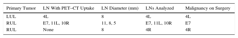
A total of 97 patients with NSCLC were identified, of whom 23 had undergone surgical resection with mediastinal lymph node dissection, and this group formed the final study cohort. Fifteen were men, and mean age was 65.49±9.8 years. Samples from 35 enlarged lymph nodes were obtained by EBUS-TBNA and thoracotomy/VATS, and results were concordant in 32: 11/12 in E7, 9/10 in 4R, 8/9 in 4L, 3/3 in 10R and 1/1 in 11L. Three false negatives were obtained, as shown in Table 1. The prevalence of mediastinal lymph node infiltration with negative EBUS-TBNA was 8.6%, with a NPV per lymph node of 91.4%. In total, 30 lymphadenopathies showed pathological uptake on PET–CT: 24 N2 (cN2) and 6 N3 (cN3), with a prevalence and NPV of 12% and 87.5%, and 0% and 100%, respectively.
Recent clinical guidelines1,2 recommend mediastinoscopy after a negative EBUS when the mediastinum is abnormal, defined as the presence of enlarged lymph nodes with pathological uptake on PET–CT, according to the conclusions of a Bayesian analysis which determined that the post-test probability of malignancy in this group of patients would be high, at around 20%.3 This estimate was made by taking into account the results of the ASTER study,4 a trial comparing EBUS-TBNA with EBUS-TBNA plus mediastinoscopy, randomized at a ratio of 1:1, which showed that the combination of both techniques was more sensitive than each one separately.
However, this conclusion is rather controversial. In our series, in patients with a moderate to high risk of N2-N3, the NPV of EBUS-TBNA is high, in line with findings from various studies in which it ranged between 89% and 99%.5–9 According with these results, the possibility has been raised that in resectable NSCLC, a negative EBUS-TBNA would not need further confirmation by mediastinoscopy, as suggested by recent guidelines from the Spanish Society of Pulmonology and Thoracic Surgery.10 This recommendation is further strengthened by evidence that mediastinoscopy is not superior to EBUS-TBNA in nodal staging, and indeed its sensitivity is similar and sometimes lower.11,12 This was also shown in the ASTER study,4 which reported that to improve sensitivity, 11 mediastinoscopies would have to be performed to obtain 1 positive case. Therefore, as the authors themselves admit, confirming all cases with negative EBUS-TBNA by mediastinoscopy might not be necessary.
Nevertheless, it would be advisable to try to identify any features that could be associated with a greater likelihood of “unexpected” nodal involvement. In this respect, our study revealed that our 3 false negatives had the common factor of a centrally located tumor, predominantly in the upper lobes. This finding has already been recognized as a predictor of malignancy in patients with negative EBUS-TBNA: Ong et al.13 showed that the presence of nodal metastases in patients with a normal mediastinum according to imaging techniques, of which 37% were detected by EBUS-TBNA, correlated significantly with central tumors, and of these, 67% were located in the upper lobes, a finding similar to that obtained in previous prospective studies.14 Similarly, Talebian Yazdi et al.,15 in a large series, found that central tumor location, along with enhanced uptake on PET, were factors predictive of false negatives in subjects with negative EBUS-TBNA.
This study has the limitations typical of a retrospective design and a small sample size, so definitive conclusions cannot be reached in certain aspects, such as the possible influence of PET uptake on EBUS-TBNA false negatives. However, the limitations of imaging studies in this regard are well known,2 and if they are taken into account, we believe that our results could be of use for identifying patients in whom it may be appropriate to perform mediastinoscopy after lymph node staging by EBUS-TBNA. We were also unable to calculate the sensitivity or the positive predictive value of the technique, because positive EBUS-TBNA results are not generally confirmed by surgery.
We conclude that in patients with potentially resectable non-small cell lung cancer, a negative preoperative EBUS-TBNA might not require confirmation by mediastinoscopy in most cases, perhaps with the exception of centrally located tumors.
Please cite this article as: Gullón Blanco JA, Villanueva Montes MÁ, Rodríguez López J, Sánchez Antuña A. Ecobroncoscopia negativa en la estadificación del carcinoma broncogénico. Arch Bronconeumol. 2017;53:646–647.


