Archivos de Bronconeumologia is a scientific journal that preferentially publishes prospective original research articles whose content is based upon results dealing with several aspects of respiratory diseases such as epidemiology, pathophysiology, clinics, surgery, and basic investigation. Other types of articles such as reviews, editorials, a few special articles of interest to the society and the editorial board, scientific letters, letters to the Editor, and clinical images are also published in the Journal. It is a monthly Journal that publishes a total of 12 issues and a few supplements, which contain articles belonging to the different sections.
All the manuscripts received in the Journal are evaluated by the Editors and sent to expert peer-review while handled by the Editor and/or an Associate Editor from the team. The Journal is published monthly in English.
Manuscripts will be submitted electronically using the following web site: https://www.editorialmanager.com/ARBR/, link which is also accessible through the main web page of Archivos de Bronconeumologia.
Access to any published article, is possible through the Journal's web page as well as from PubMed, Science Direct, and other international databases. Furthermore, the Journal is also present in Twitter and Facebook. The Journal expresses the voice of the Spanish Respiratory Society of Pulmonology and Thoracic Surgery (SEPAR) as well as that of other scientific societies such as the Latin American Thoracic Society (ALAT) and the Iberian American Association of Thoracic Surgery (AICT).
Authors are also welcome to submit their articles to the Journal's open access companion title, Open Respiratory Archives.


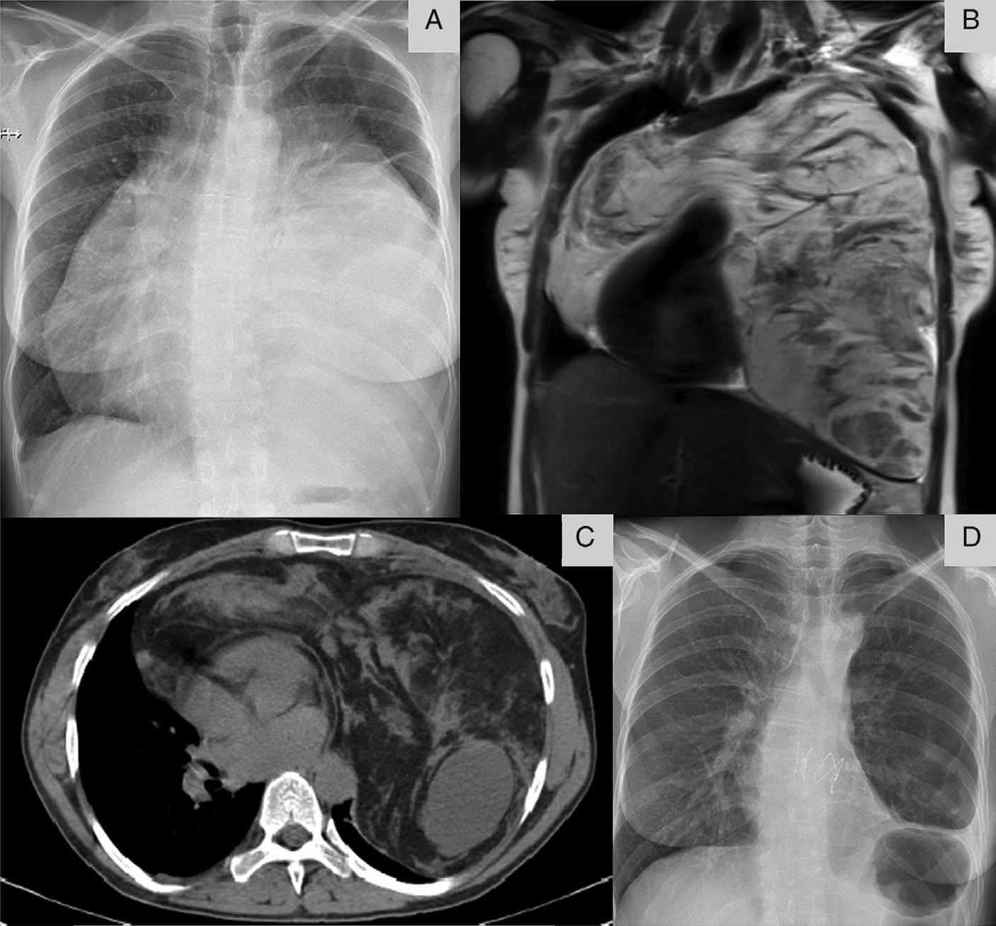
 Chest X-ray (posteroanterior) revealed a large opacity obscuring the heart border (silhouette sign). (B and C) Chest MRI and CT showing a large heterogeneous mass overlying the heart. (D) Chest X-ray (posteroanterior) after surgical excision showing lung expansion.")


