


Atrial myxomas are the most common benign primary cardiac tumors. They occur preferentially in the left atrium and clinical manifestations can be varied, with asthenia, anorexia, and weight loss due to intracavitary flow obstruction in the mitral valve and systemic embolisms; however, only exceptionally do they cause pulmonary thrombosis.1–3
A 47-year-old woman, smoker of 20 pack-years, with no other history of interest, was admitted to the respiratory medicine department with a 4-month history of dyspnea on moderate exertion, general malaise, asthenia, right pleuritic pain, and polyarthralgia. She had also had fever and mild hemoptysis in the previous 4 days. Physical examination showed basal oxygen saturation 94%, heart rate 103bpm, BP 107/73mmHg, with signs of pleural effusion on auscultation. Of note on clinical laboratory tests were elevated CRP (95.7mg/l) and 18500leukocytes/mm3 with neutrophilia. Chest X-ray showed consolidation in the left lower lobe with associated pleural effusion. Blood cultures, sputum microbiology, and antigen detection in urine were negative for pneumococcus and Legionella. Diagnostic thoracentesis was performed and was compatible with uncomplicated neutrophilic exudate. Cytology for malignant cells and microbiological cultures were negative. Despite starting empirical wide-spectrum antibiotic treatment, the patient's clinical, hemodynamic, and radiological situation worsened, with increased work of breathing, tendency to hypotension, and mildly altered level of consciousness. An angio-CT was performed, which revealed bilateral pulmonary thromboembolism (PTE), a filling defect in the left atrium, and multiple bilateral opacities consistent with pulmonary infarcts. Eco-Doppler of the lower limbs ruled out deep vein thrombosis (DVT).
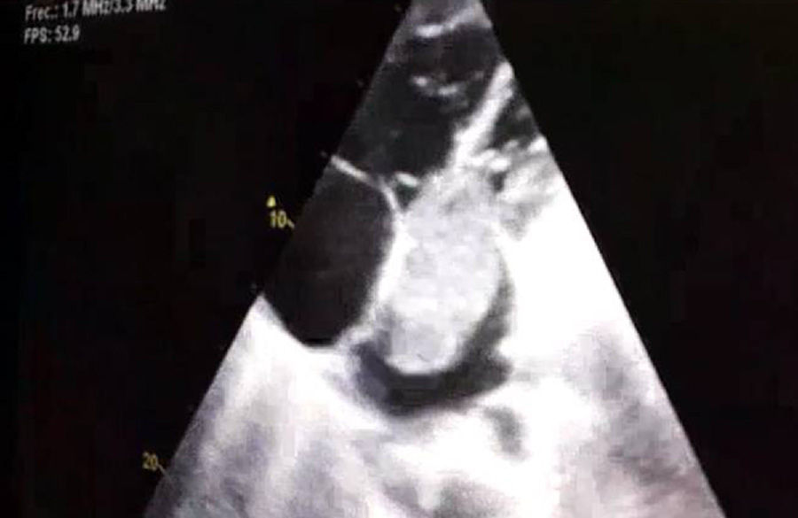
On echocardiography, a large mass was observed in the left atrium (6.3×3.2cm) that protruded in diastole toward the left ventricle, obstructing the outflow. Signs of severe pulmonary hypertension were also seen, with pulmonary artery systolic pressure 85mmHg, and estimated left ventricular ejection fraction 60% (Fig. 1).
 in diastole protruding into the left ventricle.")
In view of the severity of the clinical picture and hemodynamic instability, the patient was transferred to the Intensive Care Unit. She subsequently worsened significantly, and the cardiovascular surgery department was contacted for removal of the intracardiac tumor. The pathology report of the surgical specimen confirmed a diagnosis of atrial myxoma. After surgery and anticoagulant treatment, clinical progress was very satisfactory; in a follow-up echocardiography, the pulmonary hypertension had resolved and the pulmonary angio-CT showed resolution of the intravascular clots. A thrombophilia study was also requested, and the patient was found to be a heterozygous carrier of the factor V Leiden mutation.
A myxoma is a benign cardiac tumor; however, its benign nature is relative, since it sometimes produces distant metastases and paraneoplastic syndromes caused by cytokines and growth factors produced by the tumor.4,5 Clinical presentation depends on size and location, and it can cause intracardiac flow obstruction, stenosis, and mitral regurgitation in the left cavities.5 Echocardiography is the diagnostic test of choice, and the curative treatment is surgical resection. Being a carrier of factor V Leiden mutation is not in itself an independent risk factor for the development of PTE, but rather an additional factor along with others that confer moderate or high risk.
The presence of systemic embolisms has been frequently noted in reviews of atrial myxoma. Our case is exceptional in that the myxoma manifested as PTE without associated DVT. We believe that this may be the consequence of various concomitant risk factors: hypercoagulability derived from the myxoma, thrombophilia, concomitant infection, and obstruction of the blood flow by the tumor which may promote the presence of “in situ” thrombi in the pulmonary circulation.
Please cite this article as: Climent M, Furest I, Moragón EM. Tromboembolismo pulmonar como primera manifestación de un mixoma auricular. Arch Bronconeumol. 2018;54:46–47.


