


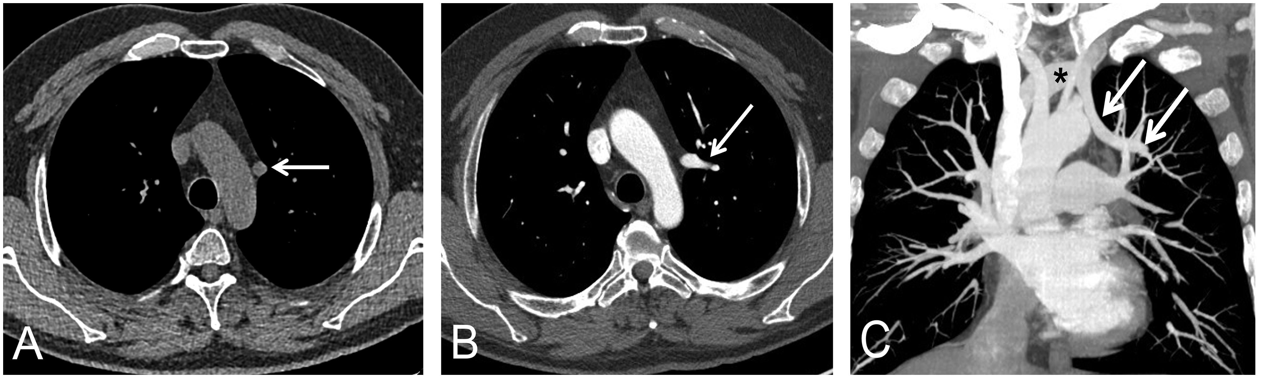
A 58-year-old man was incidentally diagnosed with a partial anomalous pulmonary venous return (PAPVR) on a baseline low-dose computed tomography (LDCT) of the thorax performed for lung cancer screening (LCS) purposes. The LDCT unexpectedly detected an anomalous vascular structure in the left mediastinum (Fig. 1A), connecting the left upper pulmonary vein with the left innominate vein, resulting in a left-to-right shunt. A contrast-enhanced chest CT demonstrated the patency of the PAPVR and ruled out other malformations (Fig. 1B and C). An echocardiography ruled out a significant hemodynamic effect of the PAPVR as well as the existence of septal defects.
 Unenhanced axial thoracic CT image (mediastinal window) shows an abnormal structure adjacent to the aortic arch (white arrow). (B) Contrast-enhanced axial CT image (mediastinal window) at a lower level shows the left upper pulmonary vein entering the mediastinum (white arrow). (C) Contrast-enhanced coronal CT maximum intensity projection image shows the left upper pulmonary vein entering the mediastinum (arrows) and connecting with the left innominate vein (asterisk).")
(A) Unenhanced axial thoracic CT image (mediastinal window) shows an abnormal structure adjacent to the aortic arch (white arrow). (B) Contrast-enhanced axial CT image (mediastinal window) at a lower level shows the left upper pulmonary vein entering the mediastinum (white arrow). (C) Contrast-enhanced coronal CT maximum intensity projection image shows the left upper pulmonary vein entering the mediastinum (arrows) and connecting with the left innominate vein (asterisk).
PAPVR is a rare congenital entity with a reported incidence of less than 1%, and less than 10% of PAPVR are left-sided. Although PAPVR is usually detected in children, it may also be diagnosed during adulthood. Up to 80% of patients with PAPVR may have an associated atrial septal defect.1 With the increasing implementation of LCS programs with LDCT, incidental thoracic findings are detected more often than before.2 Thoracic radiologists should pay attention not only to pulmonary findings on LDCT studies but also to extrapulmonary areas. To our knowledge, this is the first report of a PAPVR incidentally detected on LDCT performed for LCS.
Conflict of InterestsThe authors state that they have no conflict of interests.


