


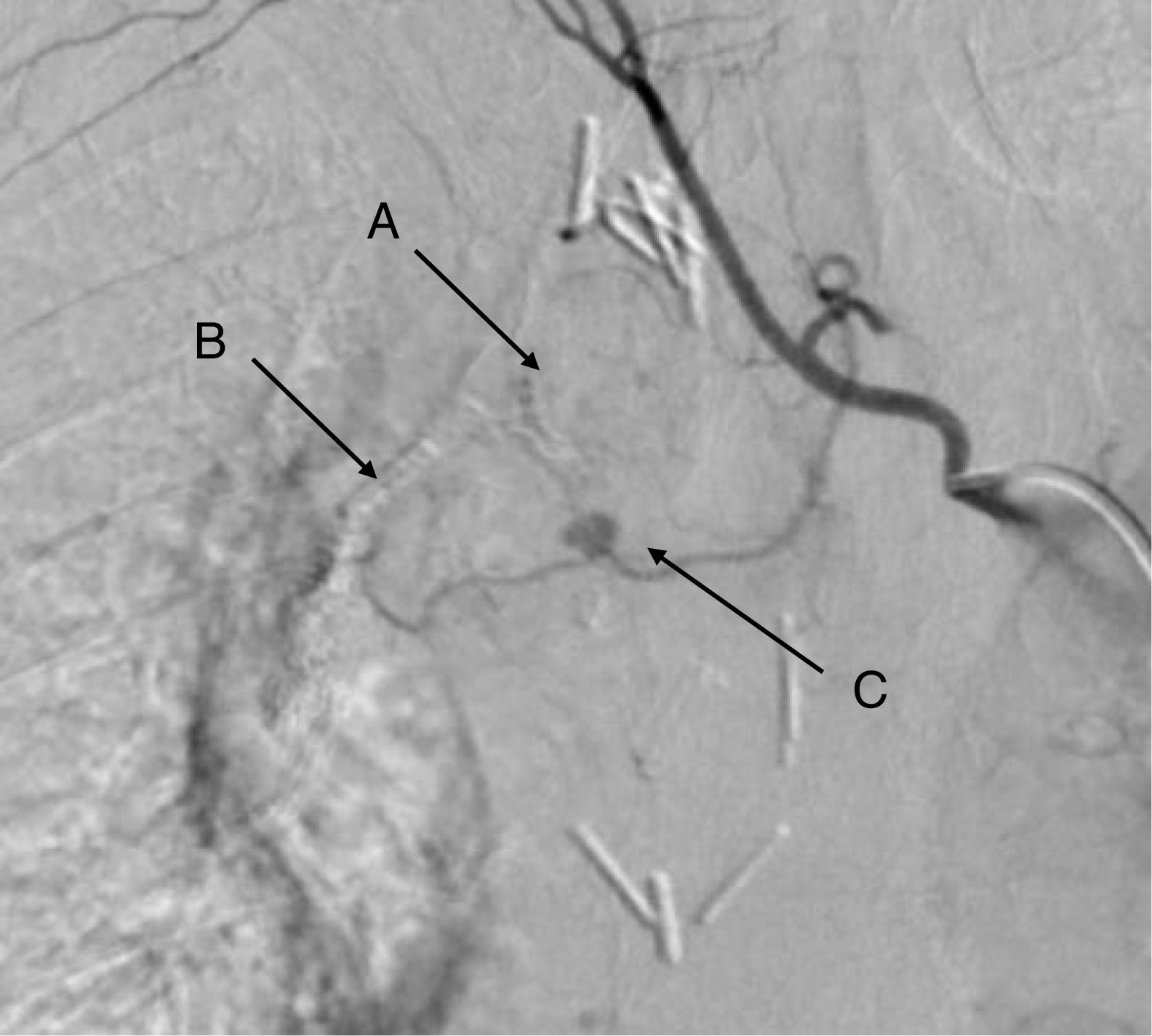
We report the case of a 65-year-old woman who underwent right upper lobectomy by video-assisted surgery. Sixty days after discharge, the patient consulted due to a 48-h history of fresh bloody sputum. Normal surgical changes were seen on chest X-ray and computed tomography (CT)-angiogram. Fiberoptic bronchoscopy was performed, which revealed mild but active bleeding from the suture of the bronchial stump. No fistula was observed. Given the endoscopic findings, pulmonary arteriography (Fig. 1) was performed, which led to the diagnosis of pseudoaneurysm of the first branch of the bronchial artery. Supraselective catherization was performed in an attempt to embolize the pseudoaneurysm, but thrombosis developed, so we opted for conservative management.
 Bronchial suture. (B) Fissure suture. (C) Pseudoaneurysm of the first branch of the bronchial artery.")
The patient has not presented hemoptysis again, and is being followed up in our hospital according to the usual protocol.
Pseudoaneurysms of the bronchial arteries are rare, and pseudoaneurysms after lobectomy are even rarer. Most cases are asymptomatic,1 although our patient developed hemoptysis due to the proximity to the stump and drainage towards the sutured bronchus. Diagnosis is made using CT angiogram or arteriography, as in our case, given the small size of the lesion. Arteriography is the most sensitive and specific diagnostic technique, and can be used to treat the pseudoaneurysm with embolization.2
Please cite this article as: Rofso Raboso P, García Fernández JL, Moreno Balsalobre R. Pseudoaneurisma de la arteria bronquial tras lobectomía superior derecha. Un caso excepcional. Arch Bronconeumol. 2019;55:48.


