


A 59-year-old male, active smoker with a history of resection of a cutaneous melanoma in the back 9 years ago. A chest X-ray was performed due to chest discomfort and the patient was sent for evaluation.
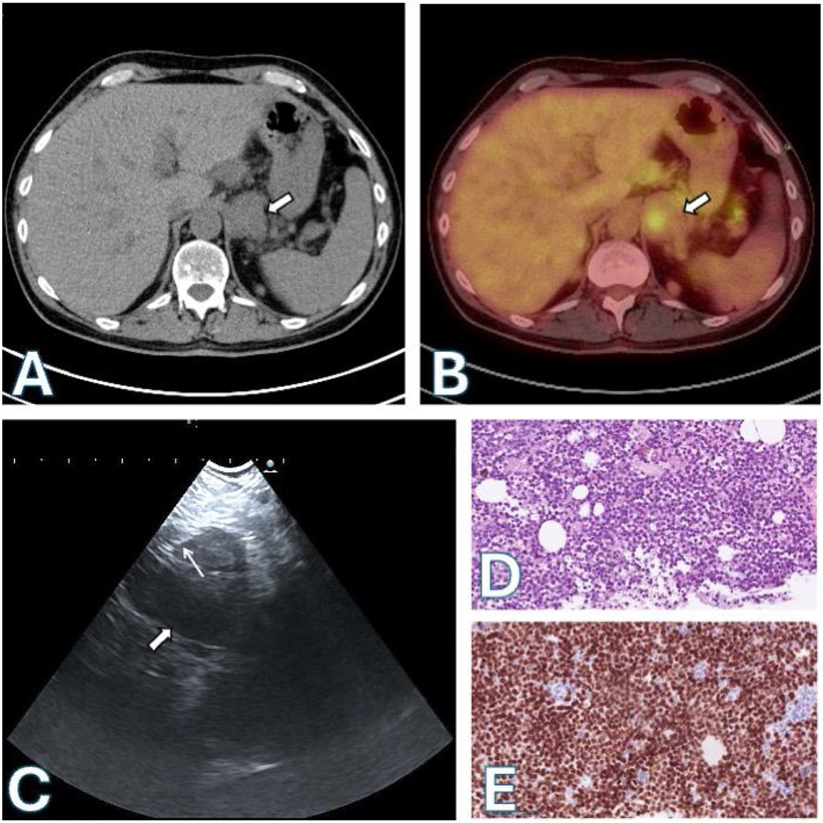
CT and PET scans revealed a pulmonary mass measuring 45mm×32mm with a Standard Uptake Volume (SUV) of 7.55. Additionally, bilateral pulmonary nodules, mediastinal lymph nodes (4R: SUV 2.24), and liver and bilateral adrenal lesions (left: 23mm, SUV 3.67) were identified.
The differential diagnosis considered the possibility of stage IV lung cancer and recurrence of melanoma, given the patient's medical history.
An echobronchoscopy (EBUS) was then performed, puncturing 4R and 11R. The procedure was followed by echoendoscopy through the esophagus with the same echobronchoscope (EUS-B) (EB19-J10U, Pentax). A transgastric fine-needle aspiration (FNA) (SonoTip EBUS Pro Flex, Medi-Globe) was performed on a left adrenal mass.
Small to medium-sized atypical cells were found in the pathologic anatomy of the adrenal gland, which were compatible with melanoma metastasis. The cells showed dual positive staining for SOX-10/Melan A. A molecular oncology test confirmed the presence of a BRAFV600E mutation. A similar finding was observed in 4R, while 11R was negative for malignancy (Fig. 1).
 and PET-CT (B) scans: left adrenal mass (thick arrows). (C) EUS-B image: transgastric needle aspiration (thin arrow) of the left adrenal mass (thick arrow). (D) Hematoxylin–eosin stain: atypical cells. (E) Positive dual SOX-10/Melan A stain.")
The adrenal glands are a frequent site of metastasis for various tumors, including up to 50% of advanced melanomas1 and around 8% of lung cancers.
EUS-B enables sampling of extrathoracic lesions and completes tumor staging by reaching areas that are not accessible by EBUS.2 Several studies have reported the efficacy of EUS-B-FNA in detecting metastases in the left adrenal gland in patients with lung cancer and other tumors. It is important to note that EUS-B-FNA is a sensitive, safe, and minimally invasive technique for providing tissue proof of left adrenal metastases.
Authors’ contributionStudy concept and design, JCH; Drafting of the manuscript, LFF, LFA; Critical revision of the manuscript for important intellectual content, JCH.
Informed consent formThe patient has signed the informed consent form.
Conflict of interestsThe authors state that they have no conflict of interests.


